Playlist
Show Playlist
Hide Playlist
Adrenal Cortex

-
Slides 05 Human Organ Systems Meyer.pdf
-
Reference List Histology.pdf
-
Download Lecture Overview
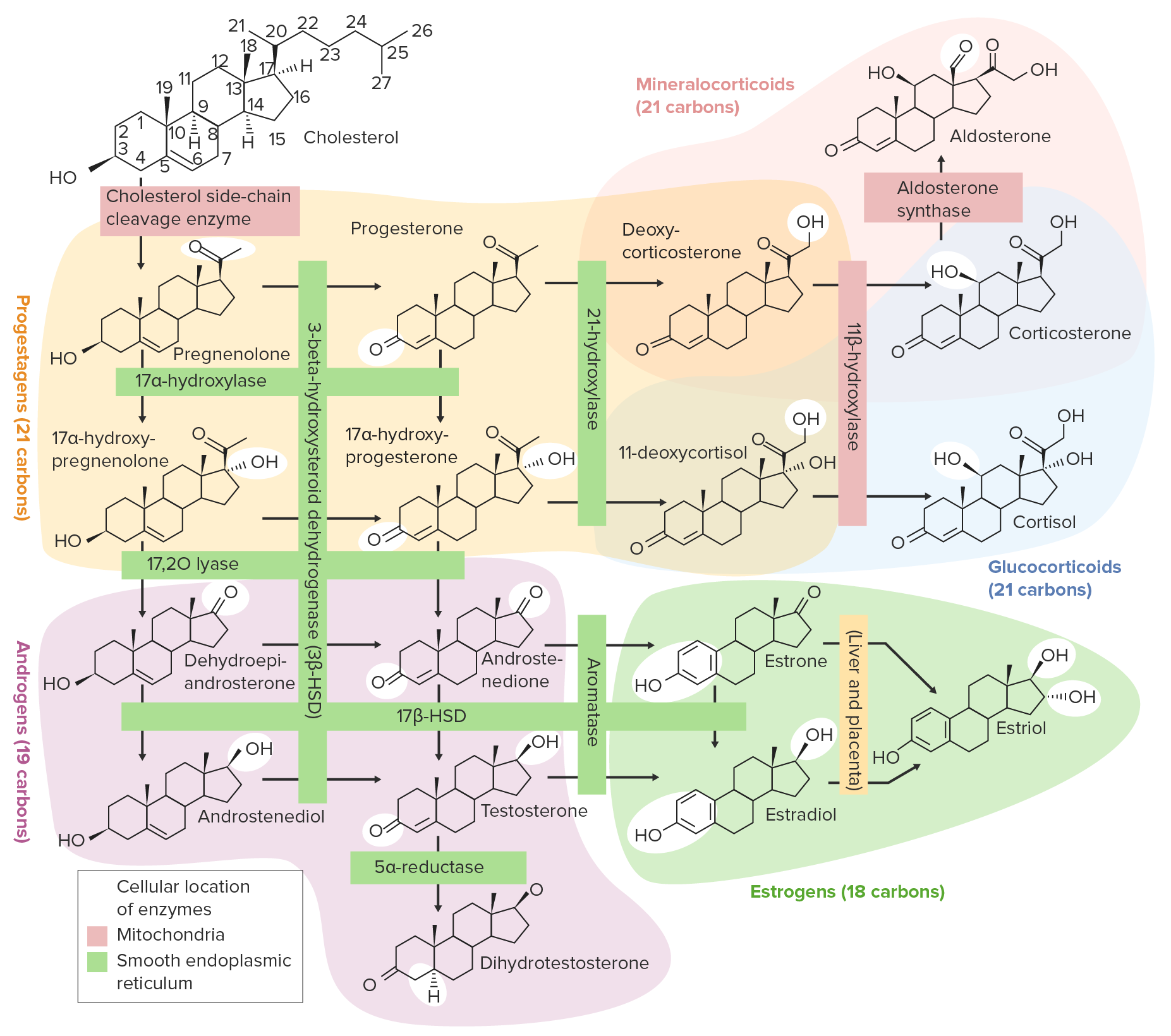
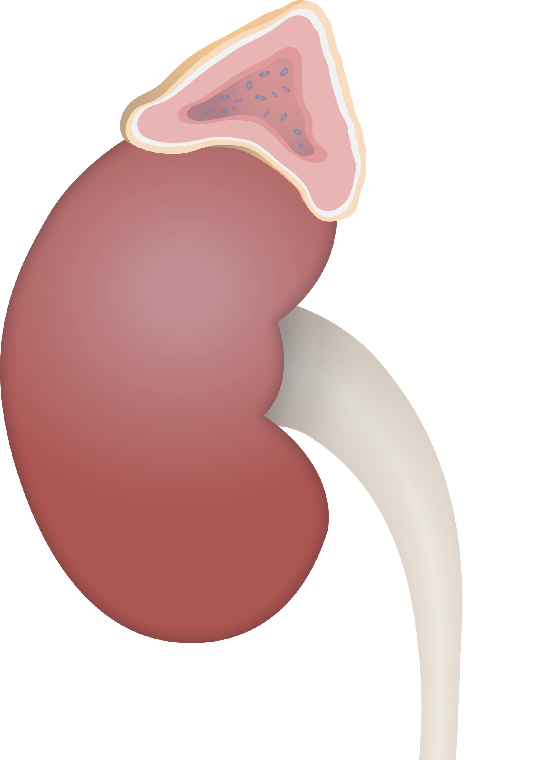
00:01 Here is in the a rather complicated slide, it’s full of a lot of information, but I’ll take it reasonably slowly and point out just the general features on this slide that are important. And then these concepts are repeated as we move through subsequent slides. First of all, in the center is the section of the cortex that I showed you on the previous slide. And those three zones I called or termed or mentioned in the previous slide, we can now name based on the histological characteristics. The outer zone near the cortex capsule, the capsular cortex is called the zonular glomerulosa for reasons I’ll mention in a moment. There is a zone called the zona fasciculata in the center, the central zone of the cortex. And towards the medulla, there is another zone, which is called the zonular reticularis. Three zones of the cortex, and down below in this section, you can see the brown stained cells making up the adrenal medulla. I’m going to talk about each of those zones in a moment. On the right-hand side, a diagrammatic representation shows you each of those three zones, as well as the medulla. And the diagram also illustrates the sort of cells that occupy each of these three zones of the cortex, and also the medulla, and the general secretory products from each of these regions. I will refer to this as we go through the remaining part of this story about the adrenal gland. I want to bring your attention though to the left-hand picture, the picture on the far left-hand side of this slide, to the left of the histological section. This is a very important concept to explain of you to understand. It’s about the blood supply to the adrenal gland. The adrenal medulla right at the bottom part of this picture with that huge round vein is the adrenomedullary vein. The adrenal medulla can get its blood from two sources. Firstly, the adrenal artery divides into small arteries in the capsule, the capsular artery, and then they in turn divide and they can send an artery, a small artery, directly into medulla to break up into a capillary bed inside the medulla, so the medulla can have a direct blood supply. 02:46 It also, as you’ll see on this diagram, it has a blood supply that comes from the adrenal cortex, that capsular artery divides into a little capillary network in the zona glomerulosa at the peripheral zone of the cortex, and then that blood percolates down along the zona fasciculata, and then around the network of cells in the zona reticularis. 03:16 And then merges with the capillary network in the medulla. So the medullary cells themselves can be affected by secretions coming from the adrenal cortex. There is this communication between the blood supply draining the cortex and the cells in the medulla. That’s a pretty important concept to understand. It’s why sometimes the adrenomedullary cells can be influenced by secretions coming from the cortex, in particular, the glucocorticoids I’ll mention in a moment. Have a look now at the zona glomerulosa. They secrete the general class of hormones called the mineralocorticoids of which aldosterone is a very important one. 04:04 If you look at the zona glomerulosa, you’ll see that the cells appear as little tiny clusters or balls of cells. Hence, the name, glomerulosa. If we go and look across the right-hand side diagram, again, it shows you a picture, a diagram of typical cells in the zona glomerulosa and the secretory or the general secretory products. I want to now describe the zona fasciculata. Have a look at the image on the left-hand side, it’s the section through that zone of the adrenal cortex, and the cells are arranged in columns. And between those columns are going to be the fine capillaries running down towards the medullary region to drain into the adrenomedullary vein. You can’t see a lot of details of these capillaries in this section, nor can you see in a lot of H&E sections of tissues and organs because they tend to collapse during processing. They live under very low blood pressures. The zona fasciculata is termed that way because, as I said, the cells are arranged into columns or fascicles. And again, you can see they’re staining differently. Some are pink and some are a whiter stain. You know, steroid hormones are produced from the adrenal cortex as they are from parts of the ovary and testes we’ll see in another lecture. Hormones have three different general classes. Some are steroid hormones, some are protein polypeptide or peptide hormones, and some are derived from amino acids. Therefore, the factories inside the cells in the organs that produce these three different sorts of organs are different, and therefore, stain different. 06:08 Here, what’s labelled is a group of cells that have little tiny droplets inside them, of cholesterol, and that’s because steroid hormones are derived initially from cholesterol, and they’re stored in these cells as these little droplets. That’s often lost during processing which in some instances like you see here the cells have a very pale appearance. 06:37 And again, on the right-hand side, it shows you an image of these cells and also a general description of the hormones produced by this fascicle of cells in the adrenal cortex, the middle layer. If you look at the zona reticularis, it’s a bit similar to the glomerulosa but it just consists of a large network of reticular epithelial type cells that are located right next to the adrenal medulla. If you look carefully in this histological section, you can see a little bit of brown stained components that represents the adrenal medulla. So the zona reticularis is the zone right next to the medulla, the inner zone of the cortex. 07:30 And again on the right-hand side, you can see a diagrammatic representation of these cells and the general products and functions of these cells in the overall role they have in the endocrine system.
About the Lecture
The lecture Adrenal Cortex by Geoffrey Meyer, PhD is from the course Endocrine Histology.
Included Quiz Questions
Glucocorticoids belong to which of the following classes of hormones?
- Steroids
- Peptides
- Glycoproteins
- Amines
- Proteins
Which of the following hormones is secreted by the zona reticularis?
- Dehydroepiandrosterone
- Cortisol
- Aldosterone
- Epinephrine
- Norepinephrine
Which of the following layers of the adrenal gland is located just beneath the capsule of the adrenal gland and contains distinct round nuclei and a higher nuclear to cytoplasmic ratio?
- Zona glomerulosa
- Zona fasciculate
- Zona reticularis
- Medulla
- Cortex
Aldosterone is secreted from which of the following parts of the adrenal gland?
- Zona glomerulosa
- Zona fasciculate
- Zona reticularis
- Medulla
- Capsule
Author of lecture Adrenal Cortex

Geoffrey Meyer, PhD
Customer reviews
5,0 of 5 stars
| 5 Stars |
|
5 |
| 4 Stars |
|
0 |
| 3 Stars |
|
0 |
| 2 Stars |
|
0 |
| 1 Star |
|
0 |
