Playlist
Show Playlist
Hide Playlist
Leukemia: Acute Leukemia – White Blood Cell Pathology

-
Slides Leukemia White Blood Cell Pathology.pdf
-
Download Lecture Overview
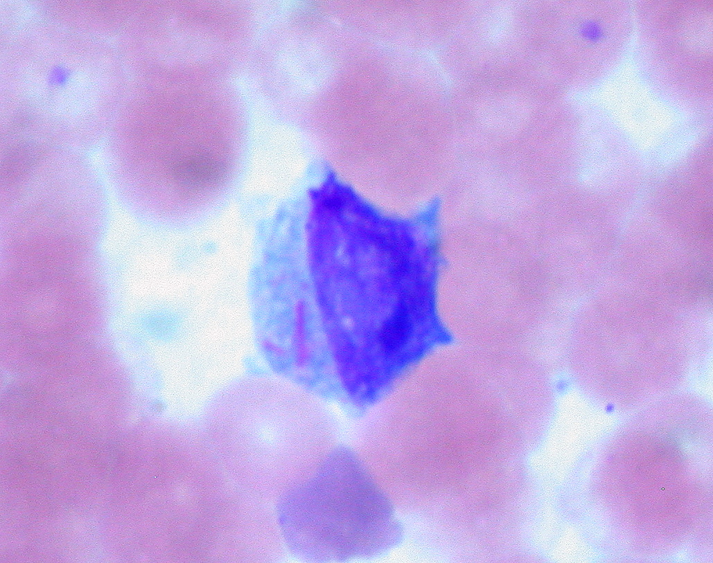
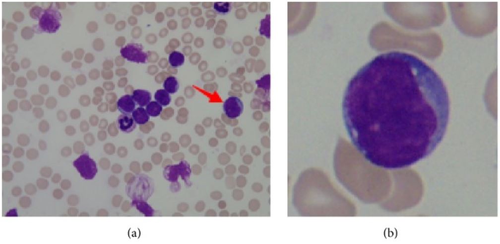
00:00 Here we’ll take a look at leukemia. 00:02 It’s a topic that most med students are quite afraid of. 00:05 But not to worry. 00:06 We will go through this together. 00:08 And by the time we’re done, your thoughts will be extremely organized and you’ll be able to identify your patient very, very confidently. 00:14 Let’s take a look. 00:17 Acute. 00:18 What does acute mean to you? It means fast. 00:22 What does leukemia mean to you? It means cancer. 00:25 Okay. 00:26 A cancer developing from where? Bone marrow. 00:30 That’s where your focus will be initially. 00:33 However at some point in time, you do know that on your peripheral blood smear you are then going to find an increased number of cells. 00:40 That puts this into leukemia. 00:42 And there is every possibility with the leukemia that these cells might then enter a lymph node. 00:47 And therefore the presentation here might be very much like a lymphoma and that will tell you as to when that will be relevant to you for symptoms and signs as far as the patient is concerned. 00:58 Is it neoplastic leukocytic origin? If you found predominance of immature cells, which are then called blasts, Where? Bone, bone, bone, bone, bone marrow. 01:10 B as in blasts. 01:12 B in bone marrow. 01:14 What is acute? What does this mean? Since this is a leukocytic type of neoplasm that the cell within the bone marrow is not being matured quick enough. 01:25 In fact, many of your cells of the neoplasm remain in its blastic form, which is a very primitive and very young cell. 01:37 By definition, ALL or AML which are both acute leukemias, by definition, you have to find greater than 20% blasts in the bone marrow. 01:49 Once that’s understood, it’s only then we can move on. 01:54 The symptoms in acute leukemia are due to bone marrow failure. 01:58 This results in decreased production of normal blood cells leading to anemia, thrombocytopenia and leukopenia. 02:06 I would like to point out that although the white blood cell count is high, these cells represent the immature, circulating blasts. 02:14 So even though there is an increase in the number of circulating leukocytes, there is functional leukopenia because these cells do not function properly. 02:23 Usually the first symptom that the patient is exhibiting would be signs and symptoms of anemia. 02:30 Meaning to say fatigue and tiredness. 02:32 The one that you’re worried about very much in terms of pancytopenia would be the susceptibility to infections, leukopenia. 02:40 Definition of acute leukemias referring to both AML or ALL, greater than 20% blasts in the bone marrow. 02:48 Etiology, chromosomal abnormalities are a possibility and Down syndrome is something we’ll take a look at. 02:57 We’ll take a look at ALL, ionizing radiation. 03:00 We have chemical exposure or maybe even perhaps alkylating agent. 03:04 Wait, hold down for a second. 03:05 You should be asking yourself what does this mean? The patient was receiving chemotherapy for another type of cancer. 03:11 And while receiving the chemotherapy unfortunately develops another type of leukemia. 03:16 So even alkylating agents that are being used to prior cancers might then unfortunately give rise to a new leukemias. 03:24 Age. 03:25 Subtypes: There will AML/ALL. 03:31 Next, what you want to do with acute leukemias? So far, you have a definition of leukemias. 03:35 This is a neoplasm of your leukocytic origin from the bone marrow. 03:43 We have greater than 20% blasts from the bone marrow. 03:46 And now, we’ll take a look at the various lineages of your cell or your bone marrow, the 2 major lineages. 03:54 One lineage will be myeloid. 03:56 The other lineage will be lymphoid. 03:58 If you’re thinking myeloid, it’s all cells except your T-cells, B-cells and natural killer cells. 04:05 So when you say acute myelogenous leukemia, you know that you’re dealing with many different types of myeloid cells. 04:13 Hence, you will be using what’s known as FAB classification, M0 all way out to M7. 04:21 By the time we come to M3 you’ve heard of, well, this is promyelocyte. 04:26 Hence, M3, which we will be focusing upon, is called your acute promyelocytic leukemia or promyelocytic leukemia. 04:35 Do not forget the other name. 04:36 By the time you’re still getting to M5, you’re producing more monocytic. 04:40 By the time you’re still getting to M5 to M6 and such, more RBC’s and M7 will be megakaryocytes. 04:47 All myeloid, all myeloid. 04:49 Disease of immature granulocytes. 04:52 Seen in, well basically, all age ranges. 04:55 Look at this, 15-60, so the age is not going to tell you much. 05:00 Tell me what you’re going to find in your bone marrow? Greater than 20% blasts in your bone marrow. 05:06 And if you’re thinking myeloid, you’re affecting all the myeloid cells except T-cells and B-cells. 05:11 What’s the other type of acute leukemia? It’s acute lymphoblastic leukemia, would be the better name that you need to know. 05:17 Once again why do we call this lymphoblastic? Because you will find greater than 20% blasts in the bone marrow. 05:23 Since we’re dealing with ALL, lympho-. 05:27 There’s only 2 types of ALL, T type and B type, B type/T type, T type/B type. 05:34 If it’s AML, there is 7 different subtypes because there are 7 different methods of developing other myeloid cells. 05:41 Clear? Next. 05:43 Disease of immature lymphocyte, pre-B or pre-T ALLs. 05:47 Typically, now you know that this is the youngest leukemia causing cancer. 05:52 So in this, you’re thinking about age group of, well, less than 15 years of age. 05:57 Number 1 leukemia in this age group.
About the Lecture
The lecture Leukemia: Acute Leukemia – White Blood Cell Pathology by Carlo Raj, MD is from the course Leukemia – White Blood Cell Pathology (WBC).
Included Quiz Questions
Which of the given values represents the minimum percentage of blast cells in acute leukemia?
- Greater than 20%
- Greater than 5%
- Greater than 15%
- Greater than 18%
- Less than 15%
Which of the following is NOT seen in bone marrow failure secondary to leukemia?
- Clock-face plasma cells
- Thrombocytopenia
- Leukopenia
- Anemia
- Pancytopenia
According to FAB classification, how many subtypes of AML are there?
- 8
- 7
- 5
- 4
- 6
Author of lecture Leukemia: Acute Leukemia – White Blood Cell Pathology

Carlo Raj, MD
Customer reviews
4,0 of 5 stars
| 5 Stars |
|
0 |
| 4 Stars |
|
1 |
| 3 Stars |
|
0 |
| 2 Stars |
|
0 |
| 1 Star |
|
0 |
I liked this video because this one is short and informative
