Playlist
Show Playlist
Hide Playlist
Acute Abdomen with Case
-
Slides Gastroenterology 19 Peritoneal Diseases.pdf
-
Reference List Gastroenterology.pdf
-
Download Lecture Overview
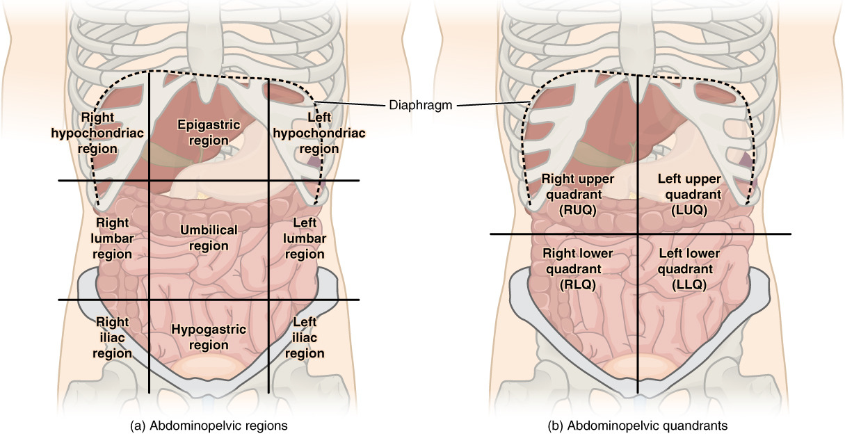
00:01 Welcome. Today we'll discuss diseases of the peritoneum. 00:04 So we'll start with a case. 00:07 A 38-year-old man with no past medical history presents to the emergency department with four hours of severe right lower quadrant abdominal pain. 00:16 The night before, he noticed a dull ache around his umbilicus. 00:20 The pain steadily worsened and is now associated with nausea and vomiting. 00:25 Vitals are notable for temperature of 39.2, heart rate 125 and blood pressure 92/50. 00:33 Physical exam reveals diffused tenderness to palpation throughout the abdomen with rebound and involuntary guarding. 00:41 Lab studies reveal a white cell count of 18,000 and lactate 3.1. 00:46 Bedside ultrasound shows peritoneal fluid in the right lower quadrant. 00:51 So, what is the best next step in management? Let's point out some key features here. 00:58 He has had periumbilical pain that then progressed to acute severe right lower quadrant pain. 01:05 He does have signs of sepsis with his temperature of 39, his tachycardia and his relative hypotension. 01:15 He also has physical exam findings that are suggestive of an acute abdomen. 01:20 In addition, his labs are somewhat concerning. 01:24 So he has a leukocytosis with an elevated lactase. 01:28 So, let's talk a bit about the acute abdomen. 01:32 The acute abdomen is what we call the condition when patients have peritonitis. 01:37 They may come in with unstable vital signs. 01:41 They will often have abdominal rigidity or involuntary guarding upon palpation of the abdomen. 01:48 They may have rebound tenderness which is when letting go of palpation causes more pain than palpation itself. 01:56 And they may have pain that worsens with any minimal movement. 02:01 So, a typical physical exam finding that I find to be most helpful is when you bump the bed that the patient is on and they -- if they have extreme pain with just that minimal movement, you can suspect that they have the acute abdomen. 02:16 It's important to remember that the acute abdomen is an emergency. 02:22 If you're concerned for any of these physical exam findings that we just described, you should emergently talk to surgery for surgical exploration. 02:31 One of the most common causes of the acute abdomen is acute appendicitis. 02:36 This happens when there is inflammation of the appendix which is a vestigial organ. 02:42 It often occurs from obstruction from a fecalith. 02:46 Patients classically present with right lower quadrant pain that begins around the umbilicus and then migrates to the right lower quadrant pain. 02:54 However, note this is not always present. 02:56 They may often have a lack of appetite or anorexia, nausea and vomiting, and they may have fevers and chills if this is a late presentation of appendicitis or if they have developed perforation. 03:09 So, there are several physical exam maneuvers that you will frequently be tested on in relation to acute appendicitis. The first is McBurney's point. 03:19 This is the point we use to describe maximal tenderness about one third the distance from the anterior superior iliac spine measured to the umbilicus. 03:30 You may also do a maneuver called Rovsing's sign. 03:35 This is when palpation of the left lower quadrant elicits pain in the patient's right lower quadrant. 03:42 Another exam maneuver you may check is the Psoas sign. 03:47 This is when you passively extend the patient's right hip and this elicits right lower quadrant pain. 03:53 Alternatively, you can do the obturator sign which is when you flex and internally rotate the patient's right hip which then elicits right lower quadrant pain. 04:05 These latter two findings work because they bring the inflamed appendix against the muscle that is named in each side and thus elicits pain in that area. 04:16 So, although you will often hear this on tests, Rovsing's, psoas and obdurator signs are actually poorly sensitive, clinically speaking when looking for acute appendicitis. 04:28 But if you happen to find one of them, they are highly specific for appendicitis, so can be helpful. 04:34 So now let's get to the diagnosis of this disease. 04:39 The diagnosis depends mostly on imaging. CT abdomen is the preferred method of diagnosis. 04:45 Here, you can see an example of a CT abdomen showing an inflamed appendix where the arrow is. 04:52 And this patient also incidentally has an abdominal aortic aneurysm as labeled by the AAA. 04:58 This is probably unrelated to this current presentation. 05:02 For those patients who -- in whom you are trying to avoid radiation exposure, you might do an ultrasound or MRI. 05:10 In addition, now, point of care ultrasound at the bedside is becoming more and more frequently used and can provide a rapid evaluation. 05:20 If you detect free fluid in the pelvis, you might suspect acute appendicitis. 05:25 So, treatment depends on whether it is perforated or non-perforated. 05:31 Once you have perforation, this is an emergency and requires exploratory laparotomy and appendectomy. 05:37 Nonperforated appendicitis can be treated with immediate appendectomy. 05:43 And in certain cases in which surgical risk is too high, you may do intravenous antibiotics and do percutaneous drainage of the abscess. 05:55 So now let's return to our case. 05:59 A 38-year-old man coming in with periumbilical pain that then progressed to severe right lower quadrant pain should prompt you to think about appendicitis. 06:09 He has signs of sepsis, so that's -- he meets SIRS criteria with a known source. 06:15 He has a physical exam signs concerning for an acute abdomen. 06:19 And his high white count with elevated lactate should prompt you to think about perforated appendicitis. 06:25 So the best next step in management would be an emergency surgical consultation for surgical exploration and washout of the abdomen.
About the Lecture
The lecture Acute Abdomen with Case by Kelley Chuang, MD is from the course Disorders of the Peritoneum.
Included Quiz Questions
Which of the following is the diagnostic test of choice for acute appendicitis?
- Abdominal CT
- Endoscopy
- Abdominal MRI
- Abdominal X-ray
- MRCP
Which of the following is the treatment of choice for nonperforated appendicitis?
- Immediate appendectomy
- Emergent exploratory laparotomy
- IV hypertonic saline
- IV anticholinergics
- Drainage of abscess
Which of the following best describes the Rovsing sign?
- Palpation of the LLQ elicits pain in the RLQ.
- Passive right hip extension elicits RLQ pain.
- Flexion and internal rotation of the right hip elicit RLQ pain.
- Palpation of the RLQ elicits pain in the LLQ.
- Passive right hip flexion elicits RLQ pain.
Customer reviews
5,0 of 5 stars
| 5 Stars |
|
5 |
| 4 Stars |
|
0 |
| 3 Stars |
|
0 |
| 2 Stars |
|
0 |
| 1 Star |
|
0 |


