Playlist
Show Playlist
Hide Playlist
Abnormalities of the Kidneys and Ureters

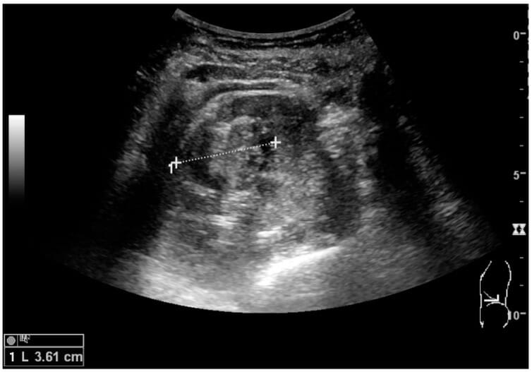

00:01 So now let's begin with the discussion on the abnormalities found within the kidneys and the ureters. 00:06 Some common renal pathology includes nephrolithiasis which is one of the most common findings found within the kidneys. 00:14 Hydronephrosis, renal cysts which are also very common incidental findings and renal cell carcinoma. So let's talk in depth about each of these abnormalities, Let's take a look at this case, this is a patient that presented asymptomatically just generalized abdominal pain. 00:32 What do you see on this image? You can see that there are multiple calcifications on both sides. 00:44 So this is an example of nephrolithiasis, seen on the abdominal plain film. 00:49 Stones can really be present anywhere within the renal collecting system, often when they're located within the ureters they can present with pain and they can result in hydronephrosis. 00:58 They're actually best evaluated on a non-contrast CT. 01:01 However, in any patient that comes in with the abdominal pain if you take a look at their KUB or their abdominal plain film you may be able to see stones, so it?s important to keep in mind to look for abnormal calcificiations. 01:13 So this is an ultrasound image of the patient that presented with right sided abdominal pain. What do you see on this image? So this is an example what renal calculi would look like on an ultrasound, they present as echogenic foci which typically shadow and again remember that shadowing is caused by calcifications on an ultrasound because the acoustic sound waves are actually reflected off of the area rather than penetrating through them, until you see an area of darkness or shadowing behind the area of calcifications. 01:48 Depending on the size of the calculus however shadowing may not always be seen so if it's a very, very small calculus it may not be, it may not have the effect of something that's a little bit larger. 01:59 So let's take a look at this case, this patient has a renal abnormality, let's take a good look at each of the images in a little more detail. 02:06 You can pause and take a good look at them and then come up with what you can find. 02:18 So the abnormality is on the right and it?s within the kidney. 02:21 So what do you think that represents? This is a coronal CT image on the same patient. 02:33 So let's take a look at this and again the arrow points to an abnormality. 02:36 So what do you think that is? This is an image, an axial CT image lower down within the pelvis. 02:47 So let me point out the anatomy here. 02:49 So this right here is the urinary bladder and that is the abnormality. 02:55 On ultrasound of the same patient we have images of both the left kidney and the right kidney. So again the abnormality is on the right. 03:02 So you can compare the two, so this is a normal left kidney and then this is the kidney with the abnormality. 03:11 So this is an example of dilatation of the renal collecting system. 03:14 It's most often due to obstruct-- an obstructing lesion within the ureter. 03:18 So in this patient the obstructing lesion is a small calculus that we saw at the ureterovesical junction, right the junction of the ureter and the bladder. 03:26 That tiny little stone that we saw in that area is causing hydronephrosis.
About the Lecture
The lecture Abnormalities of the Kidneys and Ureters by Hetal Verma, MD is from the course Abdominal Radiology.
Included Quiz Questions
Which of the following is NOT true regarding nephrolithiasis?
- Kidney stones are best evaluated on a CT with contrast.
- Kidney stones can be present anywhere in the renal collecting system.
- Stones located in the ureters can present with pain.
- On an ultrasound, renal calculi present as echogenic foci which typically shadow.
- Shadowing seen on ultrasound is a result of calcification in stones.
How do renal calculi typically present on imaging?
- Ultrasound: Echogenic foci with shadowing
- Renal calculi cannot be seen on ultrasound and require CT imaging.
- Present as solid calcifications in the kidney but cannot be seen if they have moved to the ureter
- A plain KUB X-ray can show radiolucent stones.
- On CT-KUB, stones produce a comet-tail sign.
Author of lecture Abnormalities of the Kidneys and Ureters

Hetal Verma, MD
Customer reviews
5,0 of 5 stars
| 5 Stars |
|
5 |
| 4 Stars |
|
0 |
| 3 Stars |
|
0 |
| 2 Stars |
|
0 |
| 1 Star |
|
0 |
