Playlist
Show Playlist
Hide Playlist
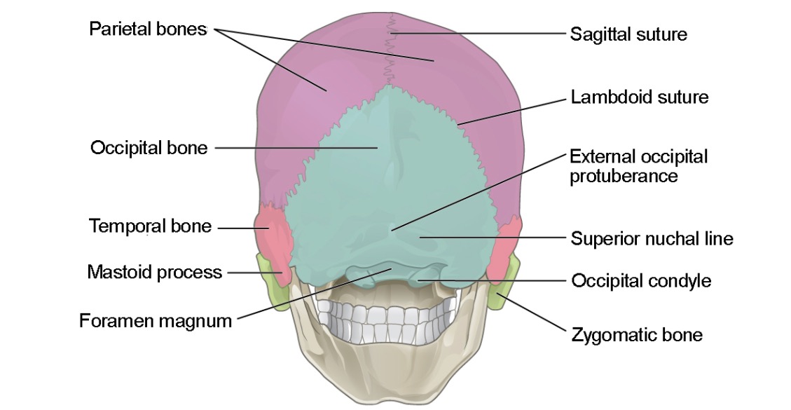
Zygomatic Bone

-
Slides Anatomy Skull.pdf
-
Download Lecture Overview
00:01 Now let's move on to the bone that is commonly known as our cheekbone, the zygomatic. 00:07 The zygomatic bone is yet another quadrangular bone, which contributes to the formation of the following structures: First, it forms a floor in the lateral wall, the ocular orbit. 00:19 Second, it contributes to the formation of the temporal and infratemporal fossae. 00:25 And lastly, it completes the zygomatic arch or what's commonly known as the cheek prominence. 00:32 Now, if you recall, we divided the frontal bone into different parts. 00:37 We have to do the same with zygomatic bone. 00:41 This bone is described as having 3 surfaces, 5 borders and 2 processes. 00:47 We will now discuss each of these parts in more detail. 00:51 Let us begin our discussion with zygomatic bone. 00:54 The first and most visible surface is the lateral facial surface. 00:59 This convex surface forms our cheek. 01:03 Here, a zygomaticofacial foramen can be found which transmits nerves and vessels of the same name. 01:10 Furthermore, the muscles of the zygomaticus major and minor also find their insertions on the facial surface of the zygomatic bone. 01:20 Behind and more medial to the facial surface is the posteromediotemporal surface. 01:26 The anterior area, the posteromedial surface is rubbed as it articulates with the zygomatic process of the maxilla. 01:34 While the posterior area, the same surface is smooth and concave. 01:39 The posterior is also the site for the opening of the zygomaticotemporal foramen. 01:45 And the last surface of the zygomatic bone is the oral surface. 01:49 This is a smooth surface which gives rise to the floor and the lateral wall of the orbital cavity. 01:56 Now let us continue our overview of the zygomatic bone by discussing its five borders. 02:02 The first of the five borders of the zygomatic bone is the anterosuperior orbital border which forms the inferolateral circumference of the orbit. 02:12 The second is the anteroinferior maxillary border through which the zygomatic articulates with the maxilla. 02:20 The third is the posterosuperior temporal border, which is L shaped and whose vertical edge is continuous with the frontal process of the zygomatic, and the horizontal edge is continuous with the zygomatic arch. 02:35 The fourth is the posteroinferior border to which the masseter muscle attaches. 02:40 And lastly, the fifth is the posteromedial border, which articulates with a greater wing of the sphenoid and the oral surface of the maxilla. 02:50 And to conclude our discussion of zygomatic, its processes have to be mentioned. 02:56 As I previously mentioned or said there are two processes. 03:00 The frontal process which articulates with the zygomatic process of the frontal bone, and with the greater wing of the sphenoid posteriorly, and the temporal process, which extends to articulate with the zygomatic process of the temporal bone. 03:17 And then this slide concludes our discussion of zygomatic bone
About the Lecture
The lecture Zygomatic Bone by Craig Canby, PhD is from the course Head and Neck Anatomy with Dr. Canby.
Included Quiz Questions
Which surface of the zygomatic bone serves as the site of insertion for the zygomaticus muscle?
- Lateral facial surface
- Posteromedial surface
- Orbital surface
- Posterolateral surface
- Inferior surface
Which of the following is an articulation of the frontal process of the zygomatic bone?
- Greater wing of the sphenoid bone
- Lesser wing of the sphenoid bone
- Temporal bone
- Orbital bone
- Lacrimal bone
Author of lecture Zygomatic Bone

Craig Canby, PhD
Customer reviews
5,0 of 5 stars
| 5 Stars |
|
5 |
| 4 Stars |
|
0 |
| 3 Stars |
|
0 |
| 2 Stars |
|
0 |
| 1 Star |
|
0 |
