Playlist
Show Playlist
Hide Playlist
Xanthogranulomatous Pyelonephritis – Complicated UTIs


-
Slides Urinary Tract Infections.pdf
-
Reference List Nephrology.pdf
-
Download Lecture Overview
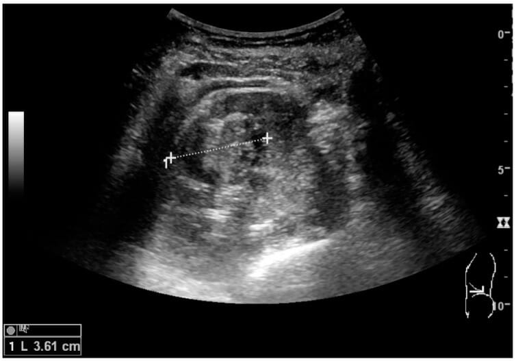
00:01 How about xanthogranulomatous pyelonephritis? This is a chronic destructive granulomatous inflammation of the renal parenchyma. 00:10 It's associated with obstruction of the urinary tract and also infected renal stones. 00:16 We see this most often in middle-aged women who have a history of UTI. 00:21 The symptoms that they're going to present with include flank pain, fever, anorexia are not being hungry and weight loss. 00:28 You can actually at times palpate a renal mass at the flank. 00:32 Diagnosis again, we're going to see pyuria on our urine analysis and a positive urine culture. 00:37 But again, this is where we want to get a CT scan of her patient. 00:41 So here we can, so a CT scan is going to demonstrate enlarge two kidneys with multilocular appearance, perhaps a presence of stones. 00:50 And then you're also going to see on CT this low-density mass that's xanthomatous tissue. 00:55 So if you look here the image on the left is actually an axial cut of a CT scan through the kidneys and look at that left kidney. 01:03 It's huge. 01:04 It's multiloculated. 01:06 This is that xanthogranulomatous inflammation, and if you look at those hypodensities there, that's the xanthomonas material. 01:14 The next image beside it is actually a sagittal section of the CT scan and once again, you can see how large that kidney is and where those xanthomatous infiltrations are. 01:23 There's also a calculus in the kidney as well. 01:26 That's that bright hyperdensity. 01:28 The microorganisms that are involved in causing xantho granulomatous pyelonephritis include E.coli, Proteus, pseudomonas aeruginosa, enterococcus, Klebsiella, and staph aureus. 01:42 The treatment once again is going to include broad-spectrum parenteral antibiotics. 01:47 Most likely patients will also need a total or at least a partial nephrectomy. 01:52 So this once again, we'll be calling our Urological colleagues in order to help us out.
About the Lecture
The lecture Xanthogranulomatous Pyelonephritis – Complicated UTIs by Amy Sussman, MD is from the course Urinary Tract Infection (UTI).
Included Quiz Questions
Which of the following is true regarding xanthogranulomatous pyelonephritis?
- It is associated with infected stones and urinary tract obstruction.
- It is characterized by acute destructive inflammation of the renal parenchyma.
- CT scan shows enlarged kidneys with high-density masses.
- Management includes broad-spectrum oral antibiotics and percutaneous drainage.
Author of lecture Xanthogranulomatous Pyelonephritis – Complicated UTIs

Amy Sussman, MD
Customer reviews
5,0 of 5 stars
| 5 Stars |
|
5 |
| 4 Stars |
|
0 |
| 3 Stars |
|
0 |
| 2 Stars |
|
0 |
| 1 Star |
|
0 |
