Playlist
Show Playlist
Hide Playlist
Warm Autoimmune Hemolytic Anemia (WAIHA): Pathogenesis

-
Slides Autoimmune Hemolytic Anemia.pdf
-
Reference List Pathology.pdf
-
Download Lecture Overview
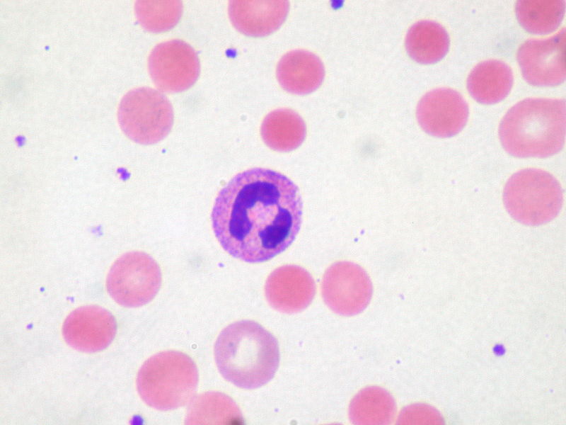
00:00 So what is warm? Warm means 37 degree Celcius. Warm reacting IgG. 00:05 Autoantibodies formed against RBC surface antigen. IgG, warm. 00:12 They're coated antibodies. Now you pay attention. 00:15 Remember that discussion that we had early on with the overview of a normocytic hemolytic anemias? And during that discussion we talked about the spleen and we talked about how the splenic macrophages were the guests. 00:28 And the guests there were having "dessert" and it was the fact that maybe a strawberry is good. 00:36 But what makes it better? You dip it into chocolate and it's coated. Right? When you coat something with, well, really sweet, whatever it might -- ingredient. 00:46 Then you're making that substance very, very attractive for? Mange. 00:53 Meaning phagocytosis. What is this chocolate that you're going to coat the RBC with? IgG. It's an Opsonin. 01:02 So now IgG warm type, you've made that RBC really, really attractive for whom? The splenic macrophages that are hungry. 01:12 So if the RBC gets killed off prematurely by the spleen, what kind of hemolysis is this? Good. 01:20 Extravascular hemolysis. Are we clear? How is your patient going to present? Significant jaundice or hemoglobinuria? Significant jaundice. 01:31 I'm gonna give you a piece of information that's really current with IGM when we get to it and you're gonna like it cuz it's a lot of integration, things you already know, it'll make perfect sense. 01:43 Let's take a look at warm. Dr. Raj, I've seen that picture before. Very good. Nice. 01:49 That picture we saw with hereditary spherocytosis. 01:52 I'm telling you ladies and gentlemen, interchangeable these are. Okay? So, in hereditary spherocytosis what were you focusing upon in this picture? The spherocyte. So therefore technically speaking you can use this and the boards will. 02:09 This picture is interchangeable with hereditary spherocytosis or warm type of autoimmune hemolytic anemia. 02:16 They're that close in imaging? Yeah, they are. They really are. 02:22 So how can you tell the difference? These will be direct Coomb's test positive. 02:28 What are you gonna find? IgG coating these RBCs. 02:33 There's no -- obviously, you know, you can't see the IgG coating the RBC. 02:37 You know, I mean if you're Superman, whatever. That's your issue. 02:42 But I'm not. Most people aren't. 02:44 So here, you can't see the IgG on the membrane. 02:47 So you have microspherocyte but this is not hereditary spherocytosis, generated with splenic macrophage removes parts of the antibody coated membrane. Clear? What's that antibody coating here? Warm. IgG. One more time. Good. 03:02 Nearly impossible to differentiate with whom? There it is, hereditary spherocytosis. 03:08 What's the test again? Direct Coomb's test. 03:10 If you missed this question, I will find a way to track you down and slap you across the face. 03:15 There's so many different times that I've told you about the differential. 03:18 Do not forget these differentials. 03:20 I'm giving these to you on purpose so that you don't confuse them on your exam.
About the Lecture
The lecture Warm Autoimmune Hemolytic Anemia (WAIHA): Pathogenesis by Carlo Raj, MD is from the course Hemolytic Anemia – Red Blood Cell Pathology (RBC).
Included Quiz Questions
Which of the following statements regarding hemolysis of RBCs in warm autoimmune hemolytic anemia is true?
- RBCs undergo extravascular hemolysis in the spleen.
- RBCs undergo extravascular hemolysis in the liver.
- RBCs undergo extravascular hemolysis in the lymph nodes.
- RBCs undergo intravascular hemolysis in small and medium blood vessels.
- RBCs undergo intravascular hemolysis in the capillary tufts of the kidney glomerulus.
The morphology of abnormal RBCs in warm autoimmune hemolytic anemia will most closely resemble that of RBCs seen in which one of the following conditions?
- Hereditary spherocytosis
- G6PD deficiency
- Hemolytic uremic syndrome
- Thalassemia
- Pyruvate kinase deficiency
Author of lecture Warm Autoimmune Hemolytic Anemia (WAIHA): Pathogenesis

Carlo Raj, MD
Customer reviews
5,0 of 5 stars
| 5 Stars |
|
1 |
| 4 Stars |
|
0 |
| 3 Stars |
|
0 |
| 2 Stars |
|
0 |
| 1 Star |
|
0 |
Great info on the d/dx for warm autoimmune hemolytic anemia and spherocytosis hereditary. This came up in a qbank for me today
