Playlist
Show Playlist
Hide Playlist
Differential Analysis of Vitamin D Dysfunction and Deficiency
-
Slides VitaminDPathologies EndocrinePathology.pdf
-
Download Lecture Overview
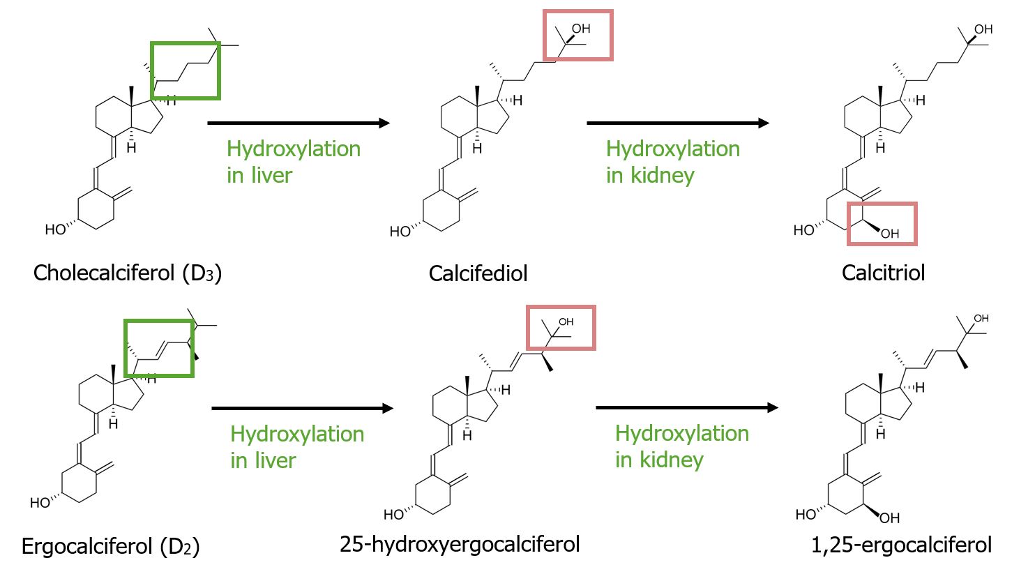
00:01 In this table, we’ll take a look at Vitamin D dysfunction. 00:03 I said dysfunction because the first column here is actual Vitamin D deficiency whereas the second column is referring to pathology of the receptors for Vitamin D and if anything, you’d find increase in Vitamin D dysfunction. 00:19 The most common cause of Vitamin D dys-deficiency in the United States, once again, is chronic renal failure. 00:25 Do not ever forget this because of the kidney dysfunction, you have decreased Vitamin D, decreased calcium, secondary hypoparathyroidism; the PTH cannot work on the kidney, thus resulting in hyperphosphatemia, hypocalcemia and may result in renal osteodystrophy. 00:44 Other cause of Vitamin D deficiency, which are quite different, would be lack of exposure to UV-rays, maybe dietary deficiency, rarely, maybe liver damage. 00:53 And those, you want to keep that separate. 00:58 Whereas if there’s resistance, then any type of resistance pathology, such as your diabetes haemolytis type 2, such as androgen-androgen insensitivity syndrome, such as your nephrogenic diabetes insipidus and in this case, we have Vitamin D and organ resistance. 01:12 You can expect the proximal hormone to then be elevated; no exception here, we have an increase in Vitamin D. 01:22 Whenever there is Vitamin D dysfunction, you can expect the calcium to be decreased; urine calcium, in fact, could be increased. 01:33 We have plasma phosphate being decreased, urine phosphate being increased. 01:43 The reason for all of this is because there isn’t enough Vitamin D. So, therefore, it’s being wasted away through the urine depleting it in your plasma. 01:56 Osteomalacia, if rickets is to a child, osteomalacia is to an adult. 02:03 So, there is no bowing of the leg, fontal bossing, rachitic rosary, the pectus carinatum and such; asymmetrical protrusion of the sternum. 02:13 Osteomalacia is in adult. 02:14 And here, if anything, we call the osteoid seam referring to that, once again, the epiphyseal plate which has been mineralized, remember that. 02:23 Osteomalacia, we’re talking about adults, mineralization is taking place, most common cause again chronic renal failure. 02:30 Usually, calcium phosphate deficiency due to dietary, malabsorption, decreased Vitamin D synthesis, that would be something like your kidney damage. 02:39 Clinical symptoms: osteomalacia, hypocalcaemia, fatigue, diffused bone pain and muscle weakness is what you’re paying attention to in osteomalacia patient, adult. 02:54 Often low or low-normal calcium, hypophosphatemia. 02:59 Now, be careful here, the reason we call this hypophosphatemia, could this ever be caused by chronic renal failure? No. 03:12 In chronic renal failure, it might result in osteomalacia, but the phosphate will be elevated. 03:22 Any other cause of Vitamin D deficiency, you can expect there to be secondary hyperparathyroidism and wasting away all the phosphate. 03:32 Mildly elevated alkaline phosphatase, once again, because of that secondary hypoparathyroidism which may then work upon that bone. 03:39 So, this first bullet point will be dealing with every type of osteomalacia due to lack of sun exposures, so on and so forth except for chronic renal failure. 03:48 That was a topic that we covered in great detail when we did nephrology. 03:53 Confirmed by bone biopsy, the tetracycline labeling showing undermineralized bone. 04:03 Both osteomalacia and rickets, you have a problem with mineralization. 04:06 In rickets, however, as a child, all kinds of issues that we discussed, osteomalacia, osteoid seam will be widened because of lack of mineralization. 04:18 Treat by repleting calcium and Vitamin D, depending as to what the cause was. 04:23 Often require quite a bit, it’s called ergocalciferol. 04:27 Here’s my dosage and timeline.
About the Lecture
The lecture Differential Analysis of Vitamin D Dysfunction and Deficiency by Carlo Raj, MD is from the course Parathyroid Gland Disorders.
Included Quiz Questions
What is the treatment for osteomalacia?
- Replete calcium and vitamin D
- Bisphosphonate therapy
- IV magnesium
- Bisphosphonate therapy and vitamin C
- Vitamin C repletion
What is NOT a characteristic of osteomalacia?
- Bowing of long bones
- Fatigue
- Diffuse bone pain
- Muscle weakness
- Decreased vitamin D synthesis
Which lab results would be expected in a patient with osteomalacia due to malabsorption?
- Normal calcium, decreased phosphate, increased alk phos, decreased vitamin D
- Increased calcium, decreased phosphate, increased alk phos, decreased vitamin D
- Normal calcium, increased phosphate, increased alk phos, decreased vitamin D
- Normal calcium, decreased phosphate, increased alk phos, increased vitamin D
- Decreased calcium, decreased phosphate, decreased alk phos, increased vitamin D
Customer reviews
5,0 of 5 stars
| 5 Stars |
|
5 |
| 4 Stars |
|
0 |
| 3 Stars |
|
0 |
| 2 Stars |
|
0 |
| 1 Star |
|
0 |


