Playlist
Show Playlist
Hide Playlist
Ventricular System: Ventricles and Communication

-
Slides 2 VentricularSystem BrainAndNervousSystem.pdf
-
Reference List Anatomy.pdf
-
Download Lecture Overview
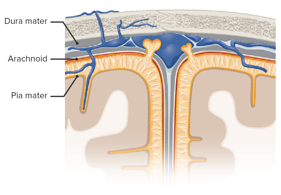
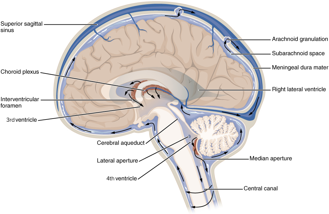
00:01 Welcome to this presentation on the ventricular system. The ventricular system is a series of fluid-filled cavities that are found in the brain. These cavities do communicate with one another. This ventricular system is in communication with the spinal cord as well. So let’s take a look at our first slide. 00:25 What I want you to understand from this slide is that the fluid-filled cavities in the brain and the fluid is cerebrospinal fluid are termed ventricles. So what are the ventricles then that contain the cerebrospinal fluid? The first to point out to you are the large, paired lateral ventricles. You can see the various components of the lateral ventricle in this particular view. It will wrap around and curve into the temporal lobe. 00:59 This extension of the lateral ventricle projects toward the occipital lobe. The next ventricular component is a single third ventricle which we see here. This has an anatomic relationship with the diencephalon. 01:21 Then your last component is the fourth ventricle that we see down in through here. It has a relationship with the brainstem lying anterior to it and then a cerebellum that lies posterior to it. These fluid-filled ventricles communicate with one another and that allows the movement or circulation of the cerebrospinal fluid from one filled cavity to another. So the components that allow for this communication then are firstly is that the lateral ventricles, each one will communicate with the third ventricle via this communication here. 02:07 This is referred to as an interventricular foramen. So there will be one on the left side as we see here. 02:14 There will be another interventricular foramen from the right lateral ventricle that would also then communicate with the third ventricle. From the third ventricle to the fourth ventricle, there is yet another opening or form of communication. This is termed the cerebral aqueduct. We see the beginning portion of it here. Then it is out of view here located behind this portion of the lateral ventricle in the temporal lobe. 02:45 Then we see it terminating in the fourth ventricle below. There are also communications between the fourth ventricle and other structures. There are two lateral apertures. Here is one. Then here is the other. 03:04 These communicate with the subarachnoid space of the brain. This space is in communication with the spinal cord, so that the cerebrospinal fluid would also flow within the subarachnoid space of the spinal cord as well as the brain. 03:21 The final communication of the fourth ventricle is with the subarachnoid space as well. That is referred to as the median aperture. The last communication that we see here is a continuation of the fourth ventricle inferiorly. This is going to communicate with the central canal of the spinal cord itself. So three apertures, two lateral, one median, and that allows the passage of cerebrospinal fluid from the fourth ventricle into the subarachnoid spaces of the brain and spinal cord. Then the central canal allows the flow of cerebrospinal fluid from the fourth ventricle into the spinal cord itself. This particular slide or table that you’re going to see is a summary of the ventricles and how they communicate with another structure. 04:28 So with the interventricular foramen, that communicates with the lateral ventricle. Then the lateral ventricles will communicate with the third ventricle. The cerebral aqueduct is going to allow for the communication of cerebrospinal fluid with the third ventricle into the fourth ventricle. You have two lateral apertures. 04:57 This will allow the movement of cerebrospinal fluid from the fourth ventricle into the subarachnoid space. 05:04 The subarachnoid space between the brain and spinal cord would be continuous. Then the median aperture of the fourth ventricle would also communicate with the subarachnoid space.
About the Lecture
The lecture Ventricular System: Ventricles and Communication by Craig Canby, PhD is from the course Ventricular System.
Included Quiz Questions
Which of the following openings does the right lateral ventricle use to communicate with the 3rd ventricle?
- Interventricular foramen
- Right cerebral aqueduct
- Left cerebral aqueduct
- Foramen of Luschka
- Foramen of Magendie
Which of the following is a continuation of the 4th ventricle inferiorly?
- Central canal
- Lateral aperture
- Interventricular foramen
- Cerebral aqueduct
- Medial aperture
Which of the following statements regarding the 4th ventricle is TRUE?
- The brain stem lies anterior to it, and the cerebellum lies posterior to it.
- It communicates with the lateral ventricles via the interventricular foramen.
- The brain stem lies posterior to it, and the cerebellum lies anterior to it,
- It communicates with the spinal cord via the cerebral aqueduct.
- It has an anatomical correlation with the diencephalon.
Through which of the following structures does CSF travel from the 4th ventricle to the subarachnoid space?
- Lateral apertures
- Central canal
- Foramen magnum
- Cerebral aqueduct
- Superior orbital fissure
Author of lecture Ventricular System: Ventricles and Communication

Craig Canby, PhD
Customer reviews
4,0 of 5 stars
| 5 Stars |
|
0 |
| 4 Stars |
|
1 |
| 3 Stars |
|
0 |
| 2 Stars |
|
0 |
| 1 Star |
|
0 |
Breakdown and delivery of the information was much better than many other lectures in this part of the curriculum. However, the lecturer seemed confused and delivered information in a confusing way towards the end. I also noticed that one of the tables in the slides changes suddenly to read something different and this confuses things further. Overall a lecture capable of delivering useful information in a concise way.
