Playlist
Show Playlist
Hide Playlist
Types of Fractures
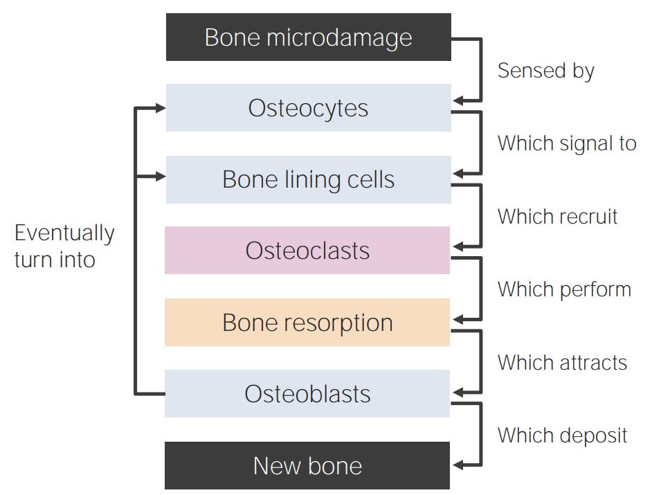
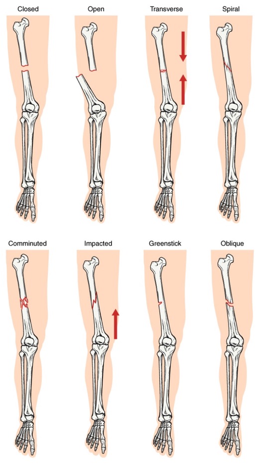
00:01 A stress fracture occurs as a result of multiple microfractures that are caused by repeated pressure on a bone. 00:08 Often, with stress fractures the radiographs are negative because these are microfractures that may not be visible radiographically but these are more easily diagnosed on an MRI or a bone scan. 00:18 So in a patient where there's a high suspicion of a stress fracture, a radiograph is usually the first line of imaging. 00:24 If that's negative because of that high clinical suspicion, the next step would be either an MRI or a bone scan. 00:30 So let's review some secondary signs of fracture. 00:35 Often the fracture line is not visible and you have to look for secondary signs to see if you think there might be a fracture present. 00:41 With a fracture you might have a joint effusion or you may have periosteal reaction or callus formation. 00:48 Periosteal reaction is actually a sign of healing but sometimes this is only the evidence in the presence of a fracture, so if you have a fracture that's not at all displaced, the fracture line may be very difficult to see and after some time as the fracture begins to heal, you'll see the surrounding callus or the periosteal reaction which will tell you that there is a healing fracture in this area. 01:08 So let's take a look at this elbow. 01:11 This is an adult patient that came in with elbow pain. 01:14 So there's a finding here posteriorly. Let's go back and take a look without the line. 01:21 So there's actually a joint effusion present here. 01:24 It's the very low density area that's seen just next to the bone. 01:30 This area right here. This can be a very subtle finding but this is only one of the signs that you could see in a radial head fracture that's the most common fracture of the elbow in an adult and the fracture itself is often very, very difficult to see. 01:45 It's the presence of that fat density posteriorly which strongly correlates with the radial head fracture. This is called the posterior fat pad sign and it's caused because the extrasynovial fat that's adjacent to the elbow is lifted away from the bone because of the presence of a joint effusion caused by the fracture. 02:03 In children, you may see the same sign and in kids, this indicates the presence of a supracondylar fracture which is the common elbow fracture found in children. 02:11 So let's take a look at this case. 02:14 This is the patient that presented with pain after fall on an outstretched hand. 02:18 This is the film of the wrist, and do you see the abnormality here? So let me point out the fracture which is right here. 02:34 This is an example of a scaphoid fracture which occurs through the waist of the scaphoid most commonly. This patient came back with a follow up, and now what do you see here? Let's take a closer look. 02:55 So the abnormality here is circled. 02:57 You can still see the lucency which represents the scaphoid fracture but do you see the subtle change in density of a portion of the scaphoid here? So the proximal pole of the scaphoid is a little bit more dense than the distal pole of the scaphoid. 03:12 This is a result of AVN or avascular necrosis of the proximal pole which occurs because the blood supply to the scaphoid runs from distal to proximal and when you have a fracture of the waist of the scaphoid, this blood supply is disrupted and results in avascular necrosis of the proximal pole. 03:29 So let's take a look at this case. 03:32 This is a patient that came in with left shoulder pain. 03:35 This is an anterior glenohumeral dislocation and it looks like there might be a fracture of the humeral head that's pointed out by the green arrow here. 03:46 So when you suspect a fracture along with a dislocation, what you wanna do is take another film after the humeral head is relocated. 03:54 So this is a post reduction film. So now, what do you see here? So the green arrow points to the abnormality. 04:09 And this is a fracture of the posterolateral aspect of the humeral head. 04:14 This is called the Hill-Sach's deformity and this is actually very commonly seen with anterior shoulder dislocation. So if you see a patient come in with anterior shoulder dislocation, you should have very low threshold for diagnosing a Hill-Sach's deformity and a post reduction film is always helpful to help you identify it. 04:31 So let's take a look at this case. 04:33 Do you see the abnormality here? This patient has a fracture. 04:39 There's a spiral fracture of the distal fibula and it extends to the distal tibiofibular joint space, so you can see here a portion of the spiral fracture which then comes around down this way and it extends right here to the distal tibiofibular joint space. 04:57 Again, when it extends to the joint space, this can have clinical implications and it's important to describe that. 05:02 So let's go back to the first case that we were looking at. 05:07 Let's take a good look. What do you think the findings are here? This is an image of the left humerus. 05:20 There's our fracture, and then what do you see surrounding the fractures? You have the fracture line right here. 05:26 Then you see an abnormality in the density of the bone around it. 05:30 This is actually an example of a pathologic fracture. 05:35 So there's a geographic lytic lesion of the humerus that has a transverse fracture through it. 05:40 This patient actually ended up having metastatic renal cell carcinoma. 05:44 So when you see a fracture, you wanna take a look at the surrounding bone as well. 05:47 If the bone looks like it's abnormal, wanna highly suspect pathologic fracture. 05:51 So we've gone over how to describe a fracture and we've gone over normal bony anatomy. 05:57 Again, fractures are something that you'll see very commonly as you come across more radiographs in the future.
About the Lecture
The lecture Types of Fractures by Hetal Verma, MD is from the course Musculoskeletal Radiology.
Included Quiz Questions
What is TRUE of a Hill Sach's deformity?
- Most commonly seen with anterior shoulder dislocation.
- Most commonly seen with posterior shoulder dislocation.
- Fracture of the anterolateral humeral head.
- Fracture of the posterolateral glenoid.
- Fracture of the medial humeral head.
Customer reviews
5,0 of 5 stars
| 5 Stars |
|
5 |
| 4 Stars |
|
0 |
| 3 Stars |
|
0 |
| 2 Stars |
|
0 |
| 1 Star |
|
0 |


