Playlist
Show Playlist
Hide Playlist
Trypanosoma brucei – Protozoa (CNS Infection)

-
Slides 13 Trypanosoma MicrobiologyAdvanced.pdf
-
Download Lecture Overview
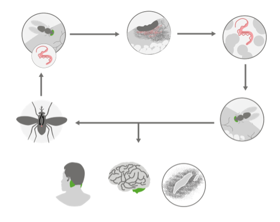
00:01 Hello and welcome to Parasites: Trypanosoma. 00:06 This is another in depth look at parasites of human importance that happened to be protozoans. 00:12 And after listening to this lecture, you'll understand the life cycles of trypanosoma parasites, and how they're transmitted to humans. 00:21 You'll be familiar with the pathogenesis of the different forms of American and African trypanosomes. 00:28 And you will know how to prevent and treat trypanosomiasis. 00:36 So, again, these are all eukaryotic protozoan parasites of medical importance. 00:43 We've looked at Plasmodia, Toxoplasma, Leishmania. 00:47 And now let's examine the trypanosomes. 00:52 Here's a photograph of the blood form of trypanosoma species. 00:57 These are very typical flagellated parasites with a lovely undulating membrane on one side. 01:04 And you can see in this photo, this is a blood smear, so there are red blood cells present. 01:09 First we'll look at the African trypanosomes. 01:12 There are African and American trypanosomes with very different outcomes. 01:18 One of the African trypanosomes, Trypanosoma brucei gambiense, let's take a look at the distribution of this. 01:27 You can see that these parasites are found largely in central Western Africa. 01:36 Very specific place, nowhere else on the planet. 01:40 Trypanosoma brucei gambiense. 01:43 The other Trypanosoma present in Africa is Trypanosoma brucei rhodesiense. 01:51 And let's take a look at the distribution of this parasite. 01:54 Look, it's very different. 01:56 It's on the Eastern Coast of South Africa. 02:00 Very different from gambiense. 02:04 These infections, these parasites, are spread by the Tsetse fly the species Glossina. 02:12 These are very large flies. 02:14 You can't tell from this photograph an inch long, that can give you quite a nasty bite and deliver quite a serious disease. 02:24 Among the African trypanosomiasis, the animal reservoirs are different for the East African and the West African parasites. 02:34 The reservoirs for the East African parasites are game animals like Impalas. 02:40 Like this animal here is an Impala and other game animals. 02:43 So these parasites exist in those reservoirs. 02:46 The West African trypanosomes have as reservoirs either humans, or domestic animals, like cattle. 02:56 So the very distinct geographic distributions of these two different African trypanosomes are a consequence of the distribution of the animal reservoir. 03:07 In both cases, they are spread by the same vector, the Tsetse fly. 03:14 Let's take a look at the pathogenesis of trypanosoma brucei. 03:19 Here's an overview. 03:20 Again, these are spread by Tsetse flies. 03:24 They inoculate the parasites, elysian forms at the side of inoculation. 03:29 They become blood parasites in the host. 03:32 And then the fly takes up parasites and repeats the cycle again. 03:37 And then there are some consequences of infection that we'll talk about. 03:42 So the infected tsetse fly is taking a blood meal, of course, that's why it's biting you. It takes a blood meal. 03:50 But in so doing it first inject saliva. 03:53 This is a common theme among the mosquitoes, and sandflies, and tsetse fly vectors that spread these diseases. 04:01 To take a blood meal, you first inject saliva, in order to give chemicals that will for example, prevent clotting of blood and in sound, injecting that saliva, there go the parasites with it. 04:14 You develop a primary lesion at the bite site. 04:18 That second circle there, which is highlighted, that is a lesion on the surface of your skin. 04:23 We call that a chancre. 04:25 The bloodstream then becomes invaded by tripomastigotes. 04:30 Those are those flagellated forms of which a photo I showed you earlier, which I think are quite beautiful. 04:37 Unfortunately, not very good for you. 04:39 They invade the bloodstream. 04:40 And of course, once they're in the blood, they can take a trip to wherever they please. 04:46 You develop a systemic illness as a consequence of the global distribution of the parasite within you with fever and lymphadenopathy. That is swollen lymph nodes. 05:00 After months, in the case of the rhodesiense or years, in the case of the gambiense, these parasites may invade the central nervous system. 05:14 So you see, here at the bottom of the right hand part of this slide you have a brain being invaded by the parasites. 05:20 An early sign that this is happening is called Winterbottom's sign, to swelling at the back of the head as you can see in this gentleman. 05:29 If you see this on someone who you know is infected, that is a bad sign, it means the parasites are likely going into the central nervous system. 05:38 And there you cannot have good consequences. 05:42 The right hand circle at the bottom there shows you a form of pathology caused by infection called perivascular cuffing when parasites are present in the blood, immune cells go around the blood vessels, and in a cross section such as this, you can see the dots are all around the blood vessel. 06:01 And that's called perivascular cuffing. 06:04 That can be used to diagnose infection. 06:07 Now, you may be understanding already at this point that this infection has gone on for months or years. 06:14 And apparently, it's not being cleared. 06:17 Well, if you thought that you'd be absolutely right, because during all these months and years of chronic infection, you have waves of parasitaemia. 06:26 So what happens is the parasites get in your blood. 06:28 We make an immune response that clears the infection, but a few are left, a few parasites are left that are not stopped by the antibody. 06:39 They have changed their coat. 06:41 And so those grow and you have another wave of parasite, you then make antibodies against those. 06:50 And of course, at this oldest time, months are going by your antibodies clear most of those are a few left who have changed their coat again. 06:57 And this goes on and on and on. 07:00 This is a hallmark of trypanosoma brucei, antigenic variation. 07:06 We can't get rid of this infection naturally. 07:09 Quite a brilliant strategy. 07:11 So here is an example of this antigenic variation. 07:15 And there is on the X axis, time and weeks. 07:18 And these are blood samples taken from an individual at different weeks after infection. 07:24 And we are measuring the number of trypanosomes in the blood on the Y axis. 07:29 You can see there's a peak and a trough. 07:32 And another peak and a trough, etc, etc. 07:35 This happens with real regularity as the parasites grow. 07:39 They're eliminated by the immune response, they change, they grow again, and so on, and so on. 07:44 This is the real problem with trying to clear this infection. 07:50 Now in the end, if you are infected, they fly, the tsetse fly can bite you and ingest blood. 07:59 And of course, take parasites up in the process and start the cycle all over again.
About the Lecture
The lecture Trypanosoma brucei – Protozoa (CNS Infection) by Vincent Racaniello, PhD is from the course Parasites.
Included Quiz Questions
Trypanosoma belongs to which variety of protozoan parasites?
- Flagellates
- Ciliates
- Sporozoans
- Sarcodines
- Non-ciliates
Which organism is the vector for Trypanosoma causing disease in humans?
- Tsetse fly
- Male Anopheles mosquito
- Sporothrix schenckii
- Ixodes scapularis
- Sandfly
What is the geographic distribution of Trypanosoma brucei gambiense?
- Central western Africa
- Central eastern Africa
- Eastern coast of South Africa
- Western coast of South Africa
- East Africa
Primary lesions caused by tsetse fly bites develop in which part of the body?
- Skin
- Stomach
- Eyes
- Liver
- Spleen
Which parasite is injected by the tsetse fly?
- Trypomastigote
- Male gametocytes
- Amastigote
- Schizont
- Merozoite
Which of the following signs or symptoms is also known as Winterbottom's sign?
- Swelling of the back of the head
- Splenomegaly
- Rhinitis
- Blackening of the skin
- Excessive lacrimation
Trypanosoma brucei causes recurrent infection by which of the following?
- Antigenic variation
- Toxin production
- Polysaccharide capsule
- IgA protease
- Haemagglutinase
Author of lecture Trypanosoma brucei – Protozoa (CNS Infection)

Vincent Racaniello, PhD
Customer reviews
5,0 of 5 stars
| 5 Stars |
|
5 |
| 4 Stars |
|
0 |
| 3 Stars |
|
0 |
| 2 Stars |
|
0 |
| 1 Star |
|
0 |
