Playlist
Show Playlist
Hide Playlist
Thymus

-
Slides 11 Human Organ Systems Meyer.pdf
-
Reference List Histology.pdf
-
Download Lecture Overview
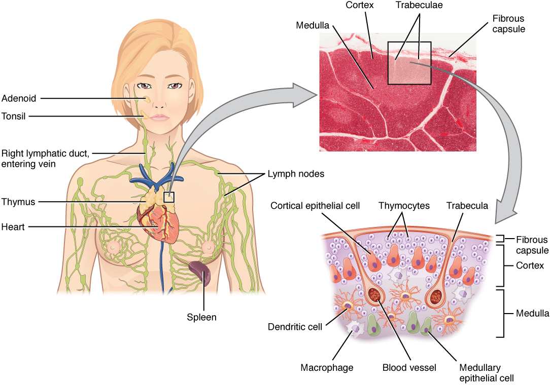
00:00 Let’s now look at the thymus. 00:02 Section here shows what the typical thymus looks like. It has got a capsule and thymic lobules. The thymic lobule has within it a dark staining cortex and a lighter staining medulla. And one feature of the thymus, one way in which you can identify the thymus from other organs is the fact that it contains thymic or Hassall’s corpuscles which I’ll show you in high magnification. The thymus is quite a complex organ, but quite easy to describe and identify histologically, as I just pointed out to you. On the left-hand side is a diagram illustrating the compartments of the thymus, the cortex and the medulla. 00:50 What essentially happens is that the cortex contains T cells undergoing education. 01:00 And they undergo education in various units or subdivisions of the cortex. And then they pass into the medulla where they undergo their final education before then moving out to the body. I sort of used the analogy that’s like a school. The cortex is like grade school or primary school where you’re receiving a certain education, and then you graduate and go to high school. Here in the cortex, T cells undergo a certain amount of training. 01:31 They go to the medulla then and then graduate as fully mature immunocompetent T cells. But one difference here is that only 2% ever graduate and move out of the thymus medulla. The rest are apoptosed or digested by macrophages. They don’t complete their education. They recognize maybe self-antigens etc. 01:55 So the body gets rid of them. On the diagram on the left, you can see rather a complex array of cells. There are six major cells that live in the thymus cortex and the thymus medulla. And they’re called epithelioreticular cells. They’re derived from the epithelium, and as I said before, they don’t secrete reticular fibres, they’re not reticular cells. The type I are the ones that are on the surface of the cortex. These are the ones that actually separate the trained or the educating thymus T cells from connective tissue. 02:41 These cells form a barrier around all the connective tissue, the capsule, the trabeculae, and even the connective tissue around the blood. So they had jobs to separate these thymus cells or T cells undergoing education. Type II cells are in the network of the cortex. 03:03 They compartmentalize the cortex into areas such as you can see shown on the diagram. 03:10 And I’m not going to go into all the details of the physiology of the thymus here, I’m just pointing out some of the histological or structural features. The type II cells are also involved in training, educating the T cells. Type III cells are also involved in education of the T cells, and they form a barrier between the cortex and the medulla, a very strong barrier to stop cells from passing from the cortex into the medulla that haven’t gone through the proper training program. The type IV cells are in the medullary side. 03:50 And what they do is they form a seal again on the medullary side so you have two cells, the type III and the type IV, making this barrier from the cortex into the medulla. 04:01 Notice that the medullaries in most of that section you see on the right-hand side, lighter staining, lighter staining because there're far less cells in there because as I said, only 2% of cells ever make it out from the thymus medulla. The rest die. And then finally, get the type VI. These are called the Hassall’s corpuscles. They’re the ones that are probably very aged epithelioreticular cells, and they form these spiral coils very easy to identify in the medulla, and therefore, they're characteristic of the thymus, and as I said earlier, enables you to identify thymus compared to other organs. And finally, we need to say something about the blood-thymus barrier. We don’t want antigens and other components of the blood, self-antigens, interfering with the education of these T cells. So, these type II and type I epithelioreticular cells form a barrier, a wrap around these capillaries. And along with the endothelial cells and the basal lamina and macrophages, prevent the contamination of the educated T cells or T cells undergoing education from being exposed to those antigens. 05:35 Macrophages live within the thymus, the cortex, and the medulla. And they are busy wrapping up or phagocytosing cells that don’t pass the grade. So again, in a review, make sure you’re aware of the structure of the cortex and the thymus medulla, and the role or function of these special epithelioreticular cells.
About the Lecture
The lecture Thymus by Geoffrey Meyer, PhD is from the course Lymphoid Histology.
Included Quiz Questions
Which of the following statements regarding regarding the thymus is INCORRECT?
- Type 2 cells compartmentalize the medulla of the thymus into different areas.
- Type 1 cells are located in the cortex of the thymus.
- Hassall's corpuscles are found in the thymic medulla.
- Type 6 cells make up Hassall's corpuscles.
- T cells undergo education in the thymic cortex.
Which of the following structures are specifically found in the thymus?
- Hassall's corpuscles
- Kupffer cells
- Trabeculae
- Mononuclear phagocyte system
- White pulp
Approximately what percent of T lymphocyte precursors undergo apoptosis?
- 98%
- 10%
- Less than 1%
- 5%
- 50%
Which types of epithelial reticular cells are mainly found in the cortex of the thymus?
- 1 and 2
- 4 and 5
- 5 and 6
- 1 and 4
- 3 and 4
The first phase of T-cell maturation occurs in which part of the thymus?
- Cortex
- Medulla
- Hassall's corpuscles
- Trabeculae
- Capsule
Which of the following types of reticular cells is primarily involved in reinforcing the blood-thymus barrier?
- 1 and 2
- 3
- 4 and 5
- 6
Author of lecture Thymus

Geoffrey Meyer, PhD
Customer reviews
5,0 of 5 stars
| 5 Stars |
|
5 |
| 4 Stars |
|
0 |
| 3 Stars |
|
0 |
| 2 Stars |
|
0 |
| 1 Star |
|
0 |
