Playlist
Show Playlist
Hide Playlist
The Structure of a Peripheral Nerve

-
Slides Cells and Basic Tissues-Nervous System.pdf
-
Reference List Histology.pdf
-
Download Lecture Overview
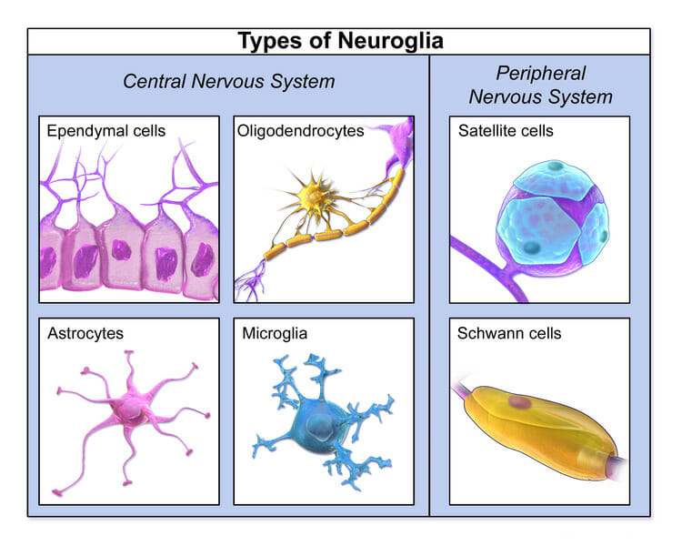
00:00 So with that background knowledge about the structure of the spinal nerve and the axons it contains, let us now have a look at how the spinal nerve is structured. It is wrapped up by a number of connective tissue components. 00:17 They're the epineurium, the perineurium and the endoneurium. The epineurium is on the outside. 00:28 The perineurium wraps around individual muscle bundles or nerve fascicles, just like when you look at the structure of muscle, individual muscle bundles are wrapped by perimysium. 00:48 Within that nerve fascicle or nerve bundle are collections of axons and each of those axons are surrounded by the endoneurium. Here is a section of a nerve cut in low magnification on the left hand side and then at higher magnification on the right hand side. And I think it is important to know how to identify these coverings when you are really looking at a real section of the nerve. Again on the outside, this rather green stained component is the epineurium. 01:32 And if you look within that, you can see a number of greenish bundle circular profiles, they represent nerve bundles or nerve fascicles. And there are number of the making up this peripheral nerve. Each of those nerve fascicles remember, is surrounded by partly the epineurium, but also the perineurium. The perineurium divides these bundles into smaller components, but also wraps up the outside of the bundle. And again let me remind you that individual axons are wrapped up by the endoneurium. It is bit hard to see that in this section at this magnification, but in the next section, it will be clearer. 02:24 Here is an image of a nerve fascicle or a nerve bundle at high magnification. 02:33 You can see on the very outside, a bright green stained component, that is collagen. It is part of the epineurium that penetrates from the outer capsule wrapping around the whole nerve and dividing the nerve into nerve bundles or nerve fascicles as you see here. Each nerve fascicle itself also has another connective tissue component called the perineurium, labelled here. It is a squamous type epithelial covering. It is a very important covering around the nerve bundle. It is a barrier. It protects the nerve fibres and nerve axons. It is fluid filled and this protection is very important to make sure the pathogens and other toxins etc do not access to the very vital structures within the nerve fascicle, the nerve axons. 03:37 And around each axon is the endoneurium. You can just see very small fibres, very small green stained components wrapping themselves around individual nerve axons. The axons shown here are represented by little dark dots and most of them have a white halo around them. 03:57 That white halo is where myelin is usually. The myelin has been lost during processing of this tissue so you do not see it. You just see the space where that myelin should be, the myelin sheaths around axons. So have a look through this image and again see if you can see little dots that represent the axons and the halo of white around those axons, you are looking there at myelinated axons. As we will see later on, some of these axons are not myelinated at all, we call them unmyelinated axons. The other nuclei you see here are other Schwann cells or they could be the nuclei of endothelial cells belonging to capillaries. 04:46 But most of the nice little circular ones you see in this section are the Schwann cells. 04:52 They are the supporting cells of the peripheral nerve axons. They are the cells that lay down in the myelin.
About the Lecture
The lecture The Structure of a Peripheral Nerve by Geoffrey Meyer, PhD is from the course Nerve Tissue.
Included Quiz Questions
Which of the following is the connective tissue covering the periphery of a nerve fascicle?
- Perineurium
- Epineurium
- Endoneurium
- Perimysium
- Epimysium
What is the outermost layer of connective tissue surrounding a peripheral nerve?
- Epineurium
- Perineurium
- Perimysium
- Endoneurium
- Epimysium
Author of lecture The Structure of a Peripheral Nerve

Geoffrey Meyer, PhD
Customer reviews
5,0 of 5 stars
| 5 Stars |
|
5 |
| 4 Stars |
|
0 |
| 3 Stars |
|
0 |
| 2 Stars |
|
0 |
| 1 Star |
|
0 |
