Playlist
Show Playlist
Hide Playlist
The Eye: General Structure
-
Slides 16 Human Organ Systems Meyer.pdf
-
Reference List Histology.pdf
-
Download Lecture Overview
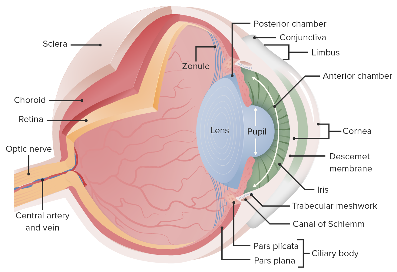
00:01 Let me just outline the general structure of the eye. On this diagram, on the right-hand side, it outlines most of the structures that I am going to describe histologically, but I want to just remind you that on the left-hand side the embryologist will explain to you in embryology lectures that the eye is actually derived from three different sources. It is developed from those components of lists that are on the left-hand side. So we are now going to look at each of those structures in turn. 00:38 I am going to start with the cornea, the front of the eye, the anterior part of the eye and go through the structures in sequence from the anterior structure of the cornea and then to the retina. 00:51 First of all, let me just outline the structures that are actually produced by the three different sources. On the left-hand side, the outer layer or tunic consists of the sclera and the cornea. The cornea is the clear transparent anterior portion of the eye. The sclera is the white portion. It is the very fibrous tuft, fibrous, outside of the eye that the extraocular muscles insert on. 01:27 The sclera in young children is quite thin and has a bluish tinge to it. As we age, it has a yellow appearance to it and it thickens. The next inner layer consists of the choroid, the stroma of the ciliary body and the iris. 01:48 This is termed the uvia. The choroid is the vascular supply to all these structures that I am going to describe. The choroids is very important because it provides support and blood supply of nutrients to the retina, which is a vascular. And then the most internal layer, the inner tunic is the retina. And the retina consists of two parts. 02:20 The neuro part which extends towards the front of the eye and sees us to be the neuro part at the ora serrata and then it extends to cover the ciliary body, ciliary processes and the iris as a double epithelial layer which I will describe later on. The ganglion cells project their axons from the retina into the optic nerve and notice in the diagram on the right-hand side that the optic nerve is actually covered by meninges, the dura, arachnoid and pia. 02:53 So we should not call it really an optic nerve, we should call it the optic tract because it is actually brain tissue. 03:06 Okay, let me start off as I said, at the front of the eye, at the cornea. The cornea is shown in low magnification on the left-hand side and then increased in magnification in the middle and also on the right-hand side. The cornea consists of two cell layers and three other noncellular layers and the thickest component is the stroma labelled here. That corneal stroma is the transparent part of our eye, the cornea. 03:43 It provides transparency and it does so because when you look at the fine structure of the stroma, it consists of about 60 layers of collagen fibrils and those layers are arranged in 19 group perpendicular to each other, layer, on layer, on layer and they're held together by glycoproteins and collagen type V and those glycoproteins, those protein glycones rather and collagen fibre actually hold all these bundles of collagen fibrils in their arrays and create this transparency. 04:19 The corneal epithelium the surface epithelium is the stratified squamous epithelium as you see in this slide. The very surface cells, the squamous cells you see at the surface have very fine microvillus projections and those projections help to maintain tears on the surface of the eye and those tears protect the eye and they also give some ultraviolet radiation protection from those cells within the cornea. Those epithelial cells do not have melamin like you see on the skin surface to protect from excessive ultraviolet radiation, but the nuclei have in them ferritin which gives that protection to the nuclei from excessive ultraviolet radiation. The corneal epithelium can replace itself very rapidly, but it does not replace itself from cells that are immediately at the base of the epithelium layer. They replace themselves by a stem cell niche or population of cells in the periphery right next to the junction of the cornea with the conjunctiva and that is to protect the cornea from conjunctival epithelium migrating its way across and being part of the cornea. The epithelium, the outer epithelium is supported by Bowman's membrane. These membranes are very thick membranes, it is a big thick basement membrane and once it is damaged it cannot regenerate. On the other side, the inner aspect of the cornea facing the anterior chamber is another basement membrane. It is a thinner basement membrane. This can repair itself on a regular basis if required and then the surface cells the inner surface of the cornea is lined by a cuboidal epithelium, the corneal endothelium it is called, unusual name endothelium really refers to the lining of the blood vessel. But here is the special exception for the cornea. Those cells are very very metabolically active. They provide nutrition and support for all the fibres that make up the cornea. The cornea is avascular. It is separated from the body, the vascular system of the body and therefore it can be used as a donation in corneal transplants. 07:06 It does not suffer from any immunological attack and that is why it can be easily donated and transferred from one person to another for a person who is in need of corneal transplant. The cornea then becomes continuous with the sclera of the eye and the junction of the epithelium with the conjunctiva. I am going to come back to this image later on because I am going to describe the canal of Schlemm, which is a very small network canal you can see at the very corner of this image, but I won't bring it to your attention yet.
About the Lecture
The lecture The Eye: General Structure by Geoffrey Meyer, PhD is from the course Sensory Histology.
Included Quiz Questions
The semitransparent sclerae in a newborn without jaundice may normally show a tint of which color?
- Blue
- Pink
- Red
- Yellow
- Green
The sclera is mainly composed of which of the following types of tissue?
- Fibrous connective tissue
- Vascular tissue
- Nervous tissue
- Cartilaginous connective tissue
- Adipose tissue
The approximate thickness of the central cornea in microns is which of the following?
- 500
- 50
- 5
- 5000
- 50,000
Customer reviews
3,0 of 5 stars
| 5 Stars |
|
1 |
| 4 Stars |
|
0 |
| 3 Stars |
|
0 |
| 2 Stars |
|
0 |
| 1 Star |
|
1 |
Histology explained really well. At each step letting us know what is going to be explained is really good.
Here is a quote from which describes Lecturio: "In order to take on this challenge, Lecturio created a high-quality digital medical education resource, which is affordable, adaptive, and personalized. We designed our platform with the needs of learners and faculty in mind, combined with the latest state-of-the-art learning technology and comprehensive monitoring and assessment features." The lecturer keeps saying that he is going to have the "anatomists" explain the structure in detail...so why are we wasting our money looking at this. I paid for a subscription to this site for "state-of-the-art learning technology." If a customer clicks on the video to see a presentation on the general structure of the eye, they shouldn't have to waste their time watching a PowerPoint with no pictures of the eye. It is an insult for customers to have this guy put up this type of presentation. My textbooks have better supplemental material .


