Playlist
Show Playlist
Hide Playlist
Testis: Structure – Male Reproductive System

-
Slides 06 Human Organ Systems Meyer.pdf
-
Reference List Histology.pdf
-
Download Lecture Overview
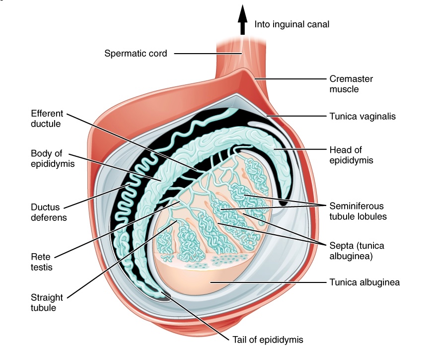
00:00 or heat gain. Now let’s look at the testis in more detail. Let’s look at its histological structure. Well, here are two images of the testis. The one on the left hand side is a histological section taken through the testis. The one on the right is a diagram I’m going to refer to, to explain some of the internal structures of the testis that you don’t really see clearly in a histological section. The outside of the testis, like any other or most other organs anyway in the body, has a capsule, a very thick dense connective tissue, collagenous capsule on the outside, called the tunica albuginea. It’s highly innervated with sensory nerve fibers. And septa, from that connective tissue capsule, penetrate into the testis and divide into a series of lobules which I’ll describe in a moment. 01:03 Within that capsule are a number of blood vessels. It’s called the tunica vasculosa. 01:09 And these blood vessels penetrate into the testis because the testis is an important endocrine gland. If you look down at the smaller section of tissues shown, that’s part of the ductus epididymis. And I point that out too because that’s the destination of the spermatozoa when they’re produced in the testis. That’s the site or the location whereby the sperm will achieve final maturity, that will become motile. Have a look at the diagram now. I’ve labelled the mediastinum. Most organs have what we call a mediastinum or a hilum where blood vessels enter and leave, or ducts or tubes enter or leave. You don’t often see it in section to the testis such as the one on the left-hand side because often, you just can’t section through that part of the testis. It’s orientated differently. In the diagram, focus your attention on the lower part of the diagram, the very dark green colored component. 02:21 It represents the capsule around the testis, the tunica albuginea that I've pointed out on the histological section. Remember, it’s collagenous, highly sensitive. Well, as I also pointed out, and you can see on the diagram, septa go into the testis. And those septa divide the testis into a number of lobules. There is up to 250 or even 300 of these separate lobules within the testis created by this connective tissue penetrating into the testis. 03:01 Within those lobules, there are very coiled tubes called seminiferous tubules. They begin in the mediastinum and they extend that in a very cold manner to the periphery of the testis, turn around and come back again. So they’re blind-ended tubes. They’re about 50 centimeters long, and there are about one to four of those in each lobule. So you can do the mathematics to work out how long this tube is going to be, these seminiferous tubules when they all join together because that represents the tube that is going to produce spermatozoa. 03:53 That tube, that seminiferous tubule, is lined by a very special epithelium. It’s called the seminiferous epithelium. And those epithelial cells are the ones that undergo differentiation to produce sperm. And that’s what we are going to examine in the next part of this lecture. 04:16 First of all, just note on the diagram that these tubes enter the mediastinum. They all come together on colorless in structures we call the rete testis. And then the spermatozoa pass from the rete testis in the mediastinum of the testis. They pass out of the testis through efferent ducts or the ductus efferentes or ductuli efferentes depending on whether you’re talking about singular or plural. And then, they then pass into the epididymis that I pointed out earlier, and that becomes continuous with the ductus deferens. All those tubular structures I’m going to talk about in a later lecture, but I think it’s very important to point out their associated with the testis at this stage. Let’s look at
About the Lecture
The lecture Testis: Structure – Male Reproductive System by Geoffrey Meyer, PhD is from the course Reproductive Histology.
Included Quiz Questions
Which ONE of the following statements is INCORRECT?
- Mature spermatozoa are stored in the seminal vesicles.
- Spermatozoa mature in the epididymis.
- Tunica albuginea is the fibrous covering of the testis.
- The tunica vasculosa is the vascular layer of the testis.
- Septae divide the testis into lobules.
What is the correct route of the spermatozoa once they are formed?
- Seminiferous tubule -> rete testis -> efferent ductule -> epididymis
- Seminiferous tubule -> efferent ductule -> rete testis -> epididymis
- Seminiferous tubule -> epididymis -> rete testis -> efferent ductule
- Epididymis -> seminiferous tubule -> rete testis -> efferent ductule
- Seminiferous tubule -> rete testis -> afferent ductule -> epididymis
Which of the following shows the correct route of mature spermatozoa during ejaculation?
- Tail of epididymis, deferent duct, spermatic cord, ejaculatory duct
- Tail of epididymis, spermatic cord, deferent duct, ejaculatory duct
- Spermatic cord, tail of epididymis, deferent duct, ejaculatory duct
- Spermatic cord, tail of epididymis, ejaculatory duct, deferent duct
- Spermatic cord, ejaculatory duct, tail of epididymis, deferent duct
Where does the sperm achieve final maturity?
- Epididymis
- Tunica albuginea
- Tunica vasculosa
- Seminiferous tubule
- Vas deferens
What is the major component of tunica albuginea?
- Collagen
- Keratin
- Elastin
- Smooth muscle
- Capillary network
Author of lecture Testis: Structure – Male Reproductive System

Geoffrey Meyer, PhD
Customer reviews
5,0 of 5 stars
| 5 Stars |
|
1 |
| 4 Stars |
|
0 |
| 3 Stars |
|
0 |
| 2 Stars |
|
0 |
| 1 Star |
|
0 |
I really appreciate the labeled pictures. I really helped to understand concepts
