Playlist
Show Playlist
Hide Playlist
Superficial and Middle Layers (AC) – Anatomy of the Forearm

-
Slides 06 UpperLimbAnatomy Pickering.pdf
-
Download Lecture Overview
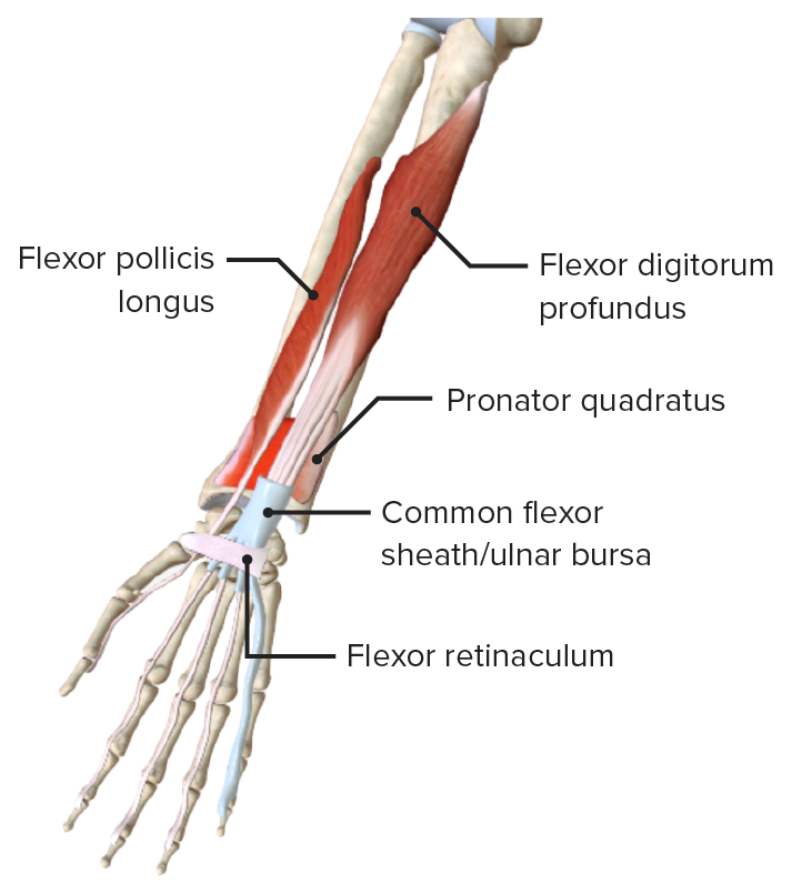
00:00 on the radio-ulnar joints. Here, we can look at the anatomy with all of these muscles put together. We can see the actual region of the forearm is quite complicated with all of these muscles. And here, we’re just looking at the superficial and the middle layers. 00:18 We can see on this superficial layer, we have those muscles, pronator teres, flexor carpi radialis, palmaris longus, and flexor carpi ulnaris. We can see they’re all radiating away from this medial epicondyle region, and they’re passing across the forearm. 00:37 So we can see pronator teres here, we can see flexor carpi radialis, and then giving rise to its long tendon. We can see palmaris longus that is passing down here towards the palm. And then we can see flexor carpi ulnaris on this most medial aspect running down here. So this is in the superficial layer. We can also see in this middle layer, we have flexor digitorum superficialis. Flexor digitorum superficialis lies deep to this superficial layer. 01:14 So to see that, we’ve just cut away a piece of flexor carpi radialis here. We can see a bit more of pronator teres running around here. And we’ve just reflected this muscle, brachioradialis. 01:26 So we’ve cut through flexor carpi radialis. We’ve cut through palmaris longus. 01:33 And underneath, we can see this large muscle belly, which is flexor digitorum superficialis. We can see here we’ve got its radial head. And here, we’ve got its humero-ulnar head. 01:47 Remember, flexor digitorum superficialis originated from the radius, the ulna, and the humerus, and we can see all of those heads. Ultimately, it was giving rise to a muscle belly. It was giving rise to four tendons that went to digits 2, 3, 4, and 5. So, as these muscles pass towards the wrist, they are held in position by the flexor retinaculum. The flexor retinaculum is a really important structure, and that it prevents bowstringing of these tendons. 02:22 So it’s held in position by flexor retinaculum, and also the palmar carpal ligament. And we’ll see this when we move into the hand. Although brachioradialis, which is the muscle we can see being reflected here, this muscle also flexes the elbow. So although brachioradialis flexes the elbow, which is similar to what these muscles do, it is actually within the posterior compartment. And it’s in the posterior compartment because it’s supplied by the radial nerve. So although we can see that muscles in the anterior compartment flex the elbow, brachioradialis, although it flexes the elbow is in the posterior compartment because of its nerve supply. Brachioradialis muscle is supplied by the radial nerve, and therefore, it’s known to be in the extensor compartment. We’ll come back to it in the next slide. If we look at the deep layers of the anterior compartment, then here we can just see the middle layers once again with these muscles haven’t been cut and brachioradialis reflected. What we’ve done now is we’ve cut through flexor digitorum superficialis. So here, it’s flexor digitorum superficialis. And on this side of the screen, we can see that we’ve cut through it. This is flexor digitorum superficialis being cut. 03:53 And what we can see is flexor digitorum profundus. Here, deep to flexor digitorum superficialis, we can see flexor digitorum profundus. We can see we also have flexor pollicis longus. 04:10 So flexor pollicis longus is running in this direction. We can see it’s coming from the radius. And also deep in here, we can just make out pronator quadratus, the deepest of the muscles. But what we can see here, these cut tendons, these are the cut tendons of flexor digitorum superficialis. And deep to them, we can see now the tendons of flexor digitorum profundus. Remembering that flexor digitorum profundus had that dual nerve supply. 04:41 The lateral muscles, the lateral tendons were innervated via the median nerve, so 2 and 3, and tendons to digits 4 and 5 were innervated via the ulnar nerve. So here we can see it quite deep.
About the Lecture
The lecture Superficial and Middle Layers (AC) – Anatomy of the Forearm by James Pickering, PhD is from the course Upper Limb Anatomy [Archive].
Included Quiz Questions
Which muscle arises from the humerus, radius, and ulna?
- Flexor digitorum superficialis
- Palmaris longus
- Flexor carpi radialis
- Flexor carpi ulnaris
- Pronator teres
Which muscle belongs to the posterior compartment of the forearm and causes flexion of the elbow joint?
- Brachioradialis
- Palmaris longus
- Flexor carpi radialis
- Flexor carpi ulnaris
- Flexor digitorum superficialis
Author of lecture Superficial and Middle Layers (AC) – Anatomy of the Forearm

James Pickering, PhD
Customer reviews
5,0 of 5 stars
| 5 Stars |
|
5 |
| 4 Stars |
|
0 |
| 3 Stars |
|
0 |
| 2 Stars |
|
0 |
| 1 Star |
|
0 |
