Playlist
Show Playlist
Hide Playlist
Structure of Peripheral Nerves and Brain

-
Slides Cells and Basic Tissues-Nervous System.pdf
-
Reference List Histology.pdf
-
Download Lecture Overview
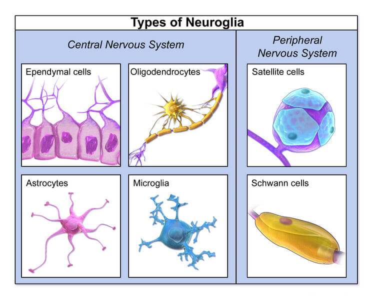
00:02 In this lecture, I would like to cover the structure of peripheral nerves and the brain. 00:09 At the end of the lecture, I would like to have some understanding of the structure of the peripheral nerve, of the basic histology of two very important components of the brain. 00:20 I am going to describe how nerve cells are supported by glial cells and also how the brain and spinal cord is protected by meninges. 00:34 The main function of the nervous system is communication and we have special sensory neurons that receive information from the periphery, from our internal organs and send their information into the brain to be processed. We have motor neurons that send information to our skeletal muscles and our smooth muscles to allow us to move individuals and for some about internal organs to change in dimension and, therefore, alter their function. 01:08 All these important occurrences have happened as a result of the neurons in the nervous system. Let us now look at the structure of a peripheral nerve. Here is a diagram showing you a section through the spinal cord. The spinal cord is in the middle of the diagram. It has two components. One is colored yellow that is the outer white matter and the internal butterfly or H-shaped structure is called the gray matter. 01:50 Concentrate on the gray matter. It has two components. It has a ventral horn and a dorsal horn on either side. The ventral horn contains the cell body of a motor neuron that is going to pass out through the ventral root and form the spinal nerve. In the dorsal horn, and in the dorsal root, are axons projecting into the central nervous system from sensory neurons. 02:31 And the cell body of these sensory neurons are located in the dorsal root ganglion. 02:40 Now that cell body also has an axon that projects out through the spinal nerve to the periphery and that axon receives information from the periphery in this case about the pain after a finger prick. And that information is traveled from the area that is being pricked by the finger through the axon all the way up through the dorsal root, through the axons associated with this sensory neuron who again as I mentioned again, the cell body is located in the dorsal root ganglion. So this is the basic structure of a spinal nerve. It consists of sensory axons carrying information into the spinal cord, and motor neurons carrying information into the skeletal muscles in the periphery. If we look across to the right-hand side of the diagram, the other half of the spinal cord there represents the visceral components. 03:46 The visceral afferent sensory neurons and the visceral efferent motor neurons, associated with the autonomic nervous system. Have a look at the tube drawn down the bottom. 04:03 It represents perhaps the wall of the gut or may be or the wall of a blood vessel. 04:12 That information is received from that structure, that internal visceral organ and the information passes through the spinal nerve into the dorsal root and into the central nervous system. 04:28 Again, the cell body of this visceral afferent sensory neuron is located in the dorsal root ganglion. All sensory neurons cell bodies are located in these dorsal root sensory ganglions, unless they are in cranial nerves. Now focus on the visceral motor components of the autonomic nervous system, and make sure you understand there are two neurons involved whereas in the somatic motor, the skeletal muscles there is only the one neuron. The preganglionic neuron originates in the lateral horn of the spinal cord. That is where the preganglionic neuron cell body is located and it then projects the axon out through the ventral root to then pass on to a ganglion and then it synapses with the postganglionic neuron in that ganglion. 05:42 And that postganglionic neuron then passes down, usually following blood vessels, to where those neurons are going to do their job where they are going to stimulates smooth muscles perhaps to contract around blood vessels or around parts of the gut. Now in this diagram, a sympathetic pathway is shown because the postganglionic cell bodies are located in here. They are located in the prevertebral ganglion, but there is also the paravertebral. 06:24 There is also the paravertebral ganglion also indicated. In sympathetic pathways, the postganglionic cell is always located in these two ganglions either released to ganglion close to the spinal cord. If this was a representation of a parasympathetic pathway, the postganglionic fibre would not be located in these ganglia, but in ganglia wire in the periphery next to the visceral organs to which they are going to send axons to innervate. And it's in those peripheral ganglia that the postganglionic fibre will originate and only travel a short distance to the site at which the activity is going to take place.
About the Lecture
The lecture Structure of Peripheral Nerves and Brain by Geoffrey Meyer, PhD is from the course Nerve Tissue.
Included Quiz Questions
The cell bodies of the sympathetic preganglionic neurons are located at which of the following sites?
- Lateral horns of the spinal cord
- Ventral horns of the spinal cord
- Dorsal horns of the spinal cord
- Dorsal columns of the spinal cord
- In ganglions near organs
Which of the following is NOT a function of glial cells?
- Impulse transmission
- Repair
- Nutrition
- Insulation
- Neuron development
Which of the following statements regarding the gray matter in the spinal cord is INCORRECT?
- It predominantly contains myelinated neurons.
- It is the H-shaped area in the middle of the spinal cord.
- It has dorsal, ventral and lateral horns.
- Cell bodies of motor neurons are present in the ventral horn.
- The dorsal horn contains sensory neurons.
Author of lecture Structure of Peripheral Nerves and Brain

Geoffrey Meyer, PhD
Customer reviews
3,0 of 5 stars
| 5 Stars |
|
1 |
| 4 Stars |
|
0 |
| 3 Stars |
|
0 |
| 2 Stars |
|
0 |
| 1 Star |
|
1 |
please use some pointers to show that part in the image. it will be easy for us to recognise it easily and it will be of great help. thank you
Very detailed inormation, helps to get good understanding, thank You!
