Playlist
Show Playlist
Hide Playlist
Sole of Foot – Anatomy of the Foot

-
Slides 07 LowerLimbAnatomy Pickering.pdf
-
Download Lecture Overview
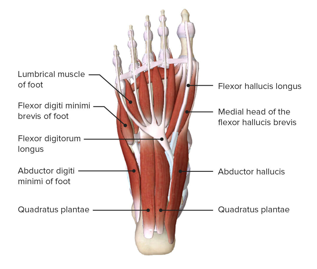
00:00 the metatarsophalangeal joints. So if we were to remove the plantar aponeurosis and we were to look deep into the foot, we’d see a whole series of those muscles, and also the nerves and the arteries. And we’ll talk about the arteries in a later lecture in more detail. But now, I just want to highlight some of the nerves. Here we’ve got the medial aspects of the sole of the foot, and here we’ve got the lateral aspect, and we can see a more radical dissection has occurred here. What we can see is that we have the lateral and the medial plantar nerves. The lateral and the medial plantar nerves are coming from the tibial nerve. And remember, the tibial nerve enter the sole of the foot posterior to the medial malleolus along with tibialis posterior, flexor digitorum longus, and flexor hallucis longus. 00:51 Remember, those tendons enter the sole of the foot posterior to the medial malleolus. 00:56 Here, we can see we’ve got the tibial nerve bifurcating into this medial plantar nerve, which we can see running along the medial aspects, and this lateral plantar nerve pass along the lateral aspect. And these are the two main nerves that are going to supply the muscles on the sole of the foot. Deeper dissection reveals some interesting blood supply. 01:20 Here, we’ve got the deep plantar arch that I spoke about previously, and it’s connecting with the lateral plantar artery that we can see here. But we’ll go over that in a later lecture. So, the sole of the foot is being divided into those compartments and those layers. But essentially, all of the muscles of the foot work together, and they work together to maintain the stunts, to maintain the arches of the foot. So, standing and making sure your balance is appropriate is carried out by these muscles of your foot, giving you a solid base. They resist the forces that want to flatten the arch of the foot so they have to hold it together, and they stabilize the foot when you are walking. So when you’re standing on a bus and it’s moving from side to side, you’ll feel your toe muscles working as you try and hold on to the sole of your foot to maintain your balance. We can see the medial and plantar nerves as a direct continuation of the tibial nerve. 02:24 And these nerves are the lateral and medial plantar arteries which are derived from the posterior tibial artery. But we’ll look at that in a later lecture. In this lecture, we looked at the dorsal aspect of the foot, intrinsic extensor muscles and extensor digitorum brevis and extensor hallucis brevis; those intrinsic extensor muscles. We looked at their relationship to the extrinsic extensor tendons. We then looked at some neurovascular relations, the dorsalis pedis and deep fibular nerve. And then we looked at the palmar aspects. We looked at the plantar fascia and the plantar aponeurosis. We then looked at the numerous compartments, the lateral, the medial, the central, and the interosseous compartments, and the various muscles that form the four layers. We then looked at the various neurovascular relations.
About the Lecture
The lecture Sole of Foot – Anatomy of the Foot by James Pickering, PhD is from the course Lower Limb Anatomy [Archive].
Included Quiz Questions
Where do the lumbricals of the foot insert?
- Medial aspects of digits 2–5
- Lateral aspects of digits 2–5
- Medial aspects of metacarpals 2–5
- Lateral aspects of metacarpals 2–5
- Posterior aspects of digits 2 - 5
Where in relation to the medial malleolus does the tibial nerve enter the foot?
- Posterior
- Anterior
- Deep
- Lateral
- Superior
Which muscle is supplied by the deep lateral plantar nerve?
- Flexor digiti minimi brevis
- Flexor hallucis brevis
- Flexor digitorum brevis
- Abductor hallucis
- First lumbricals
Author of lecture Sole of Foot – Anatomy of the Foot

James Pickering, PhD
Customer reviews
5,0 of 5 stars
| 5 Stars |
|
5 |
| 4 Stars |
|
0 |
| 3 Stars |
|
0 |
| 2 Stars |
|
0 |
| 1 Star |
|
0 |
