Playlist
Show Playlist
Hide Playlist
Small Intestine: Peyer's Patch

-
Slides 15 Human Organ Systems Meyer.pdf
-
Reference List Histology.pdf
-
Download Lecture Overview
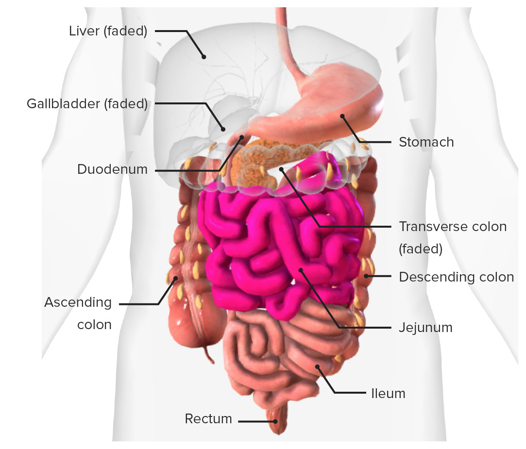
00:01 lymphatic tissue. But as I said previously, they're evidence of an immune response. 00:01 When you look at the lymphoid nodules or the lymphoid tissue you can see here, you can see a germinal center, a pale staining area. This pale staining area represents lymphocytes undergoing division originating from a lymphoblast in response to an antigen having been identified by a lymphocyte that reverts into being a lymphoblast and then producing an enormous number of lymphocytes that have the capacity to combat the invading pathogen. They differentiate into B cells and plasma cells that secrete antibodies. T cells are also present. 00:47 And I discussed the roles of these cells in a lecture on the immune system or the lymphatic tissues. Above these lymph nodules, as we call them, are M cells and other enterocytes. M cells are just a very specialized enterocyte. 01:07 They actually endocytose or ingest pathogens or antigens and then pass them out into the interstitial space beneath them where they can be readily identified by lymphocytes. And those lymphocytes then go into the lymphoid nodules next to them or in their vicinity, and they alert the immune system to mounting an immune response, and they'll return back to their location and create these nodules shown here. This slide has a diagram explaining what's happening in these Peyer's patches or lymph nodules. On the left-hand side, just make sure you review the structure of the wall of this small intestine shown here in this histological image. And on the right-hand side, there is an explanation of what's going on, and I just briefly want to summarize it. Lymphocytes travel through the blood and they can leave the blood in the various peripheral tissues by passing out of postcapillary high endothelial venules. These are very specialized epithelial cells that line these capillaries, endothelium, that actually put little flags up and attract lymphocytes into the underlying tissue when there's an inflammatory situation going on, or when there's an invading pathogen. 02:45 They sense what's going on and they alert lymphocytes to attach to their endothelial wall and then move into the system, into the tissues. 02:55 And then those lymphocytes can hunt around and detect antigens to which they're trying to detect, and then mount the response that you see here that I explained earlier. 03:06 And then they can secrete antibodies, having differentiated into plasma cells, or they can become memory cells and then spread to the rest of the body where they can then strategically locate themselves to identify future invasions of these pathogens and then react a lot quicker, or there could be T cells moving through to do their role. And they leave this area through efferent lymphatic vessels that are labelled here in yellow. There is no afferent supply, lymph supply to this area. This is where lymph vessels originate. They originate carrying lipids from lacteals in the lamina propria I'll mention in a moment. 03:59 You have seen this slide before earlier in the lecture. But the reason why I'm showing it to you now is because I want you to focus on the brush border that's in bold labelled here on the left-hand histological section. You can see it with the light microscope. 04:18 And you can also see details in the electron microscope. So I'd like to just briefly explain the importance of this brush border. On the right-hand side of this slide is an
About the Lecture
The lecture Small Intestine: Peyer's Patch by Geoffrey Meyer, PhD is from the course Gastrointestinal Histology.
Included Quiz Questions
Where are Peyer patches mainly found?
- Small intestine
- Esophagus
- Large intestine
- Liver
- Stomach
What are the aggregated lymphoid nodules in the ileum?
- Peyer patches
- Red pulp
- White pulp
- Mucosa associated lymphoid tissue
- Sinusoids
The gut-associated lymphoid tissue includes cells in which of the following tissues?
- All of the other options provided are correct.
- Lamina propria
- Intraepithelial lymphocytes
- Peyer patches
- Mesenteric lymph nodes
Author of lecture Small Intestine: Peyer's Patch

Geoffrey Meyer, PhD
Customer reviews
5,0 of 5 stars
| 5 Stars |
|
5 |
| 4 Stars |
|
0 |
| 3 Stars |
|
0 |
| 2 Stars |
|
0 |
| 1 Star |
|
0 |
