Playlist
Show Playlist
Hide Playlist
Small Intestine: Mucosal Cells

-
Slides 15 Human Organ Systems Meyer.pdf
-
Reference List Histology.pdf
-
Download Lecture Overview
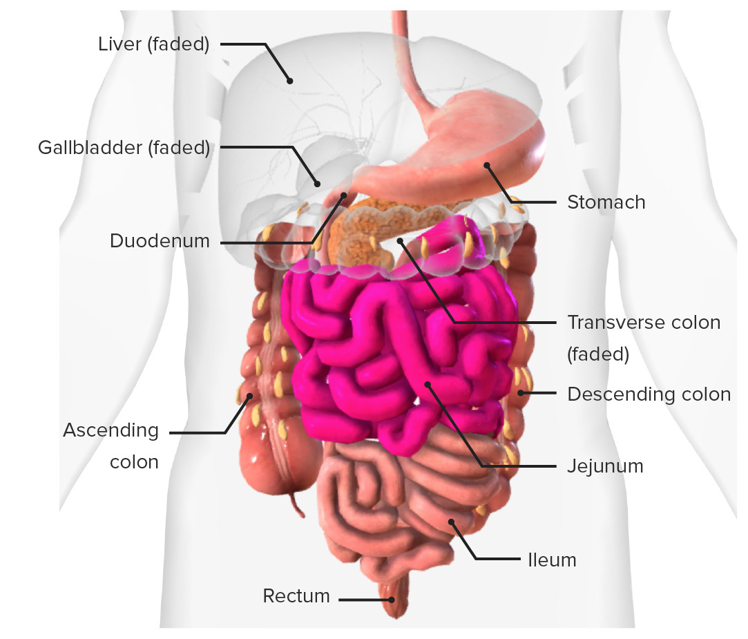
00:01 renew of epithelium here. This is now higher magnification series of three histological sections taken through the epithelial surface on the left hand side, taken at the base of the crypts or glands in the middle, and also on the right-hand side. And I want to identify various components that are important. First of all, the absorptive cell is called the enterocyte. It's specialized for absorption. It has on its surface microvilli. 00:36 When you look at the surface, you can often see a very faint pink or red line. It indicates all these microvilli. We call it a striated border or a brush border. You can see it now if you look very carefully at the apex of the epithelium. In the central section, you can see cells that contain a bright red product or content. These are called paneth cells. 01:06 And the little red granules that are stained contain antimicrobial substances that are secreted by these paneth cells into the lumen to combat these invading pathogens. Just between these glandular crypts that you see here, you can see a lot of little cells. These are the cells in the lamina propria. The lamina propria between the epithelial cells in this particular section are strongly populated by lymphocytes and cells in immune system. 01:47 On the right-hand section, you can see stem cells. As I mentioned earlier, these glands or these crypts, their secretory product really are new cells to replace ones above them that are lost at the lumen. You can see evidence of these stem cells by seeing division. You can see the metaphase, process of mitosis, clumped chromosomes on the equator of some of these cells as labelled here. And these cells will divide and finally move up along the epithelial glands and surfaces, and do their role as enterocytes, just like the other cells that have preceded them and being lost at the lumen, or they could become mucus-secreting goblet cells. 02:35 Again, on the left-hand side are the stem cells, but there's another cell that you can see here, an enteroendocrine cell. These are special endocrine cells that live on their own, embedded in the epithelium of the intestine, here in the small intestine, and indicated. 03:01 On the right-hand side, you can see first of all some goblet cells showing some nice granules of mucus inside them, but more importantly, the argentaffin cell. This is an example of an endocrine cell, an enteroendocrine cell, and endocrine cell within the gut. 03:22 Look very closely at that image or that section. I think it's a lovely section because it shows you a number of features. First of all, have a look where the lumen of this glandular component is. It's the white space in the middle. Have a look at the argentaffin cell. 03:42 You can see a round nucleus. And all those little brown granules you see are in fact the endocrine product that this cell secrete that have their effects in other parts of the gut, because they diffuse into the interstitium and into the vascular system. And that's why they're located where they are. They locate at the base of the cell, away from the apex because they're not liberated into the lumen. They're liberated into the blood stream, underneath them in the basal surface. So that's why you see them located there. 04:15 I think it's a wonderful description of these endocrine cells. 04:22 Again, let's just make sure we can see the submucosa. Previously, we are looking at the enterocytes and the crypts in the villous projections that come off the muscularis mucosa and also the glandular components in those three different sorts of folds that you see there. But make sure you are now aware of the submucosa. Contains lots of blood vessels. 04:55 You can see the bright red-stained red blood cells inside some of these vessels. And as I said before, that can move and flatten out as the gut might swell up and become full of chyme or food that's undergoing digestion. It's a very mobile layer. 05:17 Also, within that submucosa, if you look very carefully and indicated on the right-hand side, there are ganglion cells. They form the submucosal plexus or Meissner's plexus. 05:32 They're going to be postganglionic cells. They are going to be innervated by preganglionic cells that come and synapse with these postganglionic cells you see here, and they in turn send an axon, a process, and they bring about contraction of the muscularis mucosa just underneath the epithelial surfaces supporting the lamina propria as part of the mucosa. And they make the muscularis mucosa that contract independently of the muscle contraction of the muscularis externa. The muscularis externa is really only involved with peristaltic activity to move the food along the gut tube. The muscularis mucosa rather contracts and relaxes to bring about the movement of the villi, of the mucosa, to try and increase the chance of food mixing and coming in contact with the absorptive surface of the enterocytes. 06:40 And here is a description or at least an identification of the ganglion cells in the myenteric plexus which is controlling the activity, the contractile activity of the muscularis externa. 06:57 It's called the myenteric or Auerbach's plexus. And again, you see ganglion cells supported by glial cells, supportive cells. The small intestine has three different components,
About the Lecture
The lecture Small Intestine: Mucosal Cells by Geoffrey Meyer, PhD is from the course Gastrointestinal Histology.
Included Quiz Questions
Which of the following types of cells are also known as intestinal absorptive cells?
- Enterocytes
- Goblet cells
- Paneth cells
- Argentaffin cells
- Acini
Which of the following cells in the intestine constantly replenish epithelial cells that die and are lost from the villi?
- Stem cells
- Goblet cells
- Paneth cells
- Glial cells
- Enterocytes
Which of the following cells in the intestine mainly secrete mucus?
- Goblet cells
- Stem cells
- Paneth cells
- Glial cells
- Mesangial cells
Which of the following plexuses is present in the submucosa of the intestinal tract?
- Meissner plexus
- Myenteric plexus
- Lumbosacral plexus
- Kiesselbach plexus
- None of the other answer options is correct
Which of the following intestinal cells secrete antimicrobial peptides and proteins?
- Paneth cells
- Goblet cells
- Argentaffin cells
- Stem cells
- Mesangial cells
Author of lecture Small Intestine: Mucosal Cells

Geoffrey Meyer, PhD
Customer reviews
5,0 of 5 stars
| 5 Stars |
|
5 |
| 4 Stars |
|
0 |
| 3 Stars |
|
0 |
| 2 Stars |
|
0 |
| 1 Star |
|
0 |
