Playlist
Show Playlist
Hide Playlist
Small Intestine: Histology

-
Slides 15 Human Organ Systems Meyer.pdf
-
Reference List Histology.pdf
-
Download Lecture Overview
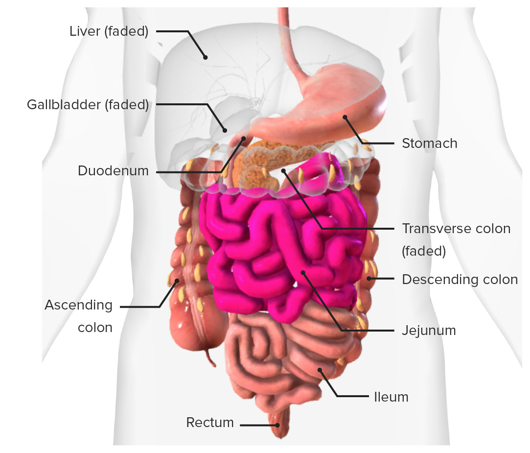
00:00 them, and alert the immune system. Starting with the small intestine, I want to just show you this slide and make sure that you fully understand the wall of the gut. The gut wall was explained in another lecture on the esophagus and stomach, but I just want to review it here. Basically, the wall of the gut has got the internal component called the mucosa that lies right against the lumen. Here, on this slide, I show you three sections. One is the diagram of the small intestine. The centre one is taken at low magnifications of the small intestine, mucosa. And on the right-hand side, you see a higher magnification. And these really illustrate the very specialized mucosa, the mucosa of the gut consists of the epithelium, the underlying lamina propria, and also the muscularis mucosa. You can see the muscularis mucosa as a thin reddish stained line of smooth muscle at the base of that section in the middle of this slide. 01:29 Focus your attention now on the left-hand diagram. On that diagram, you can see folds. 01:35 They're labelled plicae circulares. These are permanent folds in the small intestine. 01:43 They begin about six centimeters into the duodenum, and they are very prominent throughout the duodenum, and also the jejunum, and then they start to get lesser and lesser obvious at the lower end of the ileum. Have a look at the diagram and notice that they actually have a central core of connective tissue called the submucosa. The submucosa is a connective tissue layer underneath the mucosa that gives the gut mucosa mobility and flexibility. 02:26 The mucosa you see is moving all the time. As food passes along and as food is broken down in that lumen, the mucosa is continually moving about by contraction of muscularis mucosa. So that submucosa underneath the mucosa needs to be able to take in that movement. 02:53 It needs to be flexible to accommodate the moving mucosa adjacent to the lumen. In here, though, it's a permanent component of the small intestine wall. And then underneath the submucosa, you have the muscularis externa shown here on the left-hand diagram by those colored lines. They represent layers of the muscularis externa I'll describe later on through this lecture. Now look at the central slide. Make sure you can identify the submucosa in this section. Again, these are permanent folds. And on the surface of each of these submucosal projections, you can see the mucosa. And even at the low magnification you see in the centre image, you can see little tiny fine fingerlike projections coming off the core of the submucosa. They're called villi. And if you move across now and focus on the right-hand section, these villi are fingerlike projections. They're lined by epithelial cells. They're lined mainly by epithelial cells that will absorb all the nutrients and other components that are broken down in the lumen of the gut immediately adjacent to this epithelium. Small intestine doesn't have secretory cells that secrete digestive enzymes, or at least many. All the digestive enzymes come from the pancreas. Those villi have a core of lamina propria, the supporting connective tissue of the epithelium. And then underneath those little villi, there will be the muscularis mucosa that can contract and move that mucosa around. And as you can recall, underneath that then is the submucosa. We're going to look at the details of these villi in a moment. But these fingerlike projections are designed to increase the surface area of the small intestine, to increase the surface area for very effective absorption of products from the lumen across the epithelial surface into the lamina propria where there are many many blood capillaries, and also lymphatic vessels. 05:35 The epithelial cells lining these villi also invaginate deep into the lamina propria. 05:41 We call those crypts. They're essentially glands. But as we'll see, their main products are stem cells replacing the epithelial cells that are lost into the lumen, there's continual renew of epithelium here. This is now higher magnification series of three histological
About the Lecture
The lecture Small Intestine: Histology by Geoffrey Meyer, PhD is from the course Gastrointestinal Histology.
Included Quiz Questions
What are the permanent folds in the small intestine?
- Plicae circulares
- Lingula
- Linea alba
- Linea samilunaris
- Rugae
What are the small, finger-like projections of the mucosa in the small intestine?
- Villi
- Plicae
- Flagella
- Cilia
- Rugae
Plicae circulares are best developed in which section of the intestine?
- Jejunum
- Stomach
- Ileum
- Cecum
- Ascending colon
Which of the following secretes digestive enzymes?
- Pancreas
- Liver
- Spleen
- Ascending colon
- Cecum
Author of lecture Small Intestine: Histology

Geoffrey Meyer, PhD
Customer reviews
5,0 of 5 stars
| 5 Stars |
|
1 |
| 4 Stars |
|
0 |
| 3 Stars |
|
0 |
| 2 Stars |
|
0 |
| 1 Star |
|
0 |
His lectures are always amazing, simple and so clear! Thank you
