Playlist
Show Playlist
Hide Playlist
Skin (Integumentary System)

-
Slides 05 Types of Tissues Meyer.pdf
-
Reference List Histology.pdf
-
Download Lecture Overview
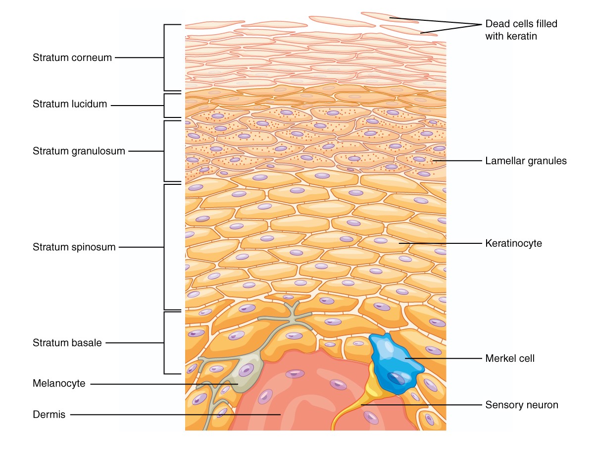
00:01 In this lecture, I am going to describe skin. It is often referred to as the integumentary system. The integument is a system of the body because it contains all the basic tissues and that is why we call it a system. We will see later on during this lecture, that skin contains epithelium and glands derived from epithelium, the exocrine components. 00:30 They contain connective tissue particularly the dermis of skin that also contains muscle and nerves. So that is why we call it a system. Now, during this lecture, I would like you to assemble information that will allow you to then answer all the questions that I list here in the learning outcomes. I am not going to go through them all here, but I will go through each of these in the course of this lecture. 01:04 Skin has got many many functions and during this lecture I am going to explain the structural components of skin that enable these functions to be fulfilled. First of all, let's make sure we can understand the layers of skin. In this section, on the right-hand side, there is an image of a section through thick skin. There are two main different types of skin, thick skin and thin skin, and really the term is a little bit confusing. Here is an image of thick skin because the epidermis, a layer I am going to describe in a moment is very very thick compared to thin skin where it is much much thinner. Here is the epidermis. 02:01 It is the surface layer. It is an epithelium. It is a very special epithelium. It is a stratified squamous epithelium because there are many many cell layers making up the epithelium as we will see in a moment. But the layer on top, that pale purple thick layer happens to be keratin and that is a very important component of skin that I will describe later on. Now this thick skin is found on the palms of our hands or the soles of our feet where we have a lot of wear and tear. But really as I said before, the term is a little bit confusing. Because really the thickest part of our skin is on our back because of the thick underlying layer, the dermis indicated here. The thinnest part of our skin on our body is the eyelid or perhaps the scrotum. In both those areas, the eyelid and the back, the epidermis is probably the same thickness. So as I said the term is a little bit confusing. 03:13 During this lecture, when I refer to thick skin, I am talking about skin on the palms of our hands and the soles of our feet. On other occasions, I would refer to thin skin. 03:25 But it is easy to describe the structure of skin looking first of all at this place of thick skin. You know sometimes we call skin either hairy or non hairy. That is another way in which we can classify skin and it is probably an easier one to understand. 03:45 We certainly do not have hair on the palms of our hand or the soles of our feet. So the two main layers indicated here, are the epidermis, the epithelium is derived from ectoderm and the underlying dermis, which is derived from mesoderm. The dermis is dense connective tissue. 04:07 When you look at it, it is very pink stained because it is full of connective tissue fibres and that dermis supports the epithelium, makes our skin very strong. Underneath the skin, underneath the dermis, you can see a very clear layer. That is the hypodermis. 04:30 It contains adipose tissue or lots and lots of fat cells. And if you look very very carefully in that hypodermis, you can make out some little round circular profiles stained bluish. 04:46 These are sections through sweat glands, one of the skin appendages I will talk about later on. They're all derived from the epidermis and during development, they grow down and become very specialized in areas of the dermis and as you see here in areas of the hypodermis. 05:10 Well, let's concentrate first of all on the epidermis. And as you see listed on the left-hand side of the slide, there are five main layers and I am going to go through these layers in some detail now. First of all, let us look at the stratum basale. It is often called the stratum germativum because it is a germinating layer. It is a single layer of cells, a single layer epithelial cells tightly bound to the underlying connective tissue, the underlying dermis by very very large numbers of hemidesmosomes. These are macular adherens special bonding structures that bind epithelial cells to the underlying connective tissue. Well the nuclei of the stratum basale lie very close together, and that's because there's not a lot of cytoplasm associated with the cells. These cells divide constantly because skin is continually renewed. 06:21 They divide constantly and as they divide, they move towards the surface to form the other layers of skin. The stratum spinosum is a thicker layer. It consists of several layers of cells. It is often called the prickle cell layer or the spinous cell layer, and that's because if you look very very carefully in this section, better still if you had the opportunity to look at this layer under higher magnification, you will see between the cells, there is a fine little clear space. And if you look carefully into that clear space, you will see these little tiny spike looking processes. These represent an artifact. In actual fact, in reality, the cells of this layer, the stratum spinosum, are linked together by long cell processes like my fingers here interlocking. And the fingers, my fingers here join to each other by very strong desmosomes. So skin, the cell layer, the stratum spinosum, the cell processes are joined together from one cell to another by very strong desmosomes join the process together, like desmosomes are holding my fingers together. It is very hard for me to separate my fingers apart because of those junctions. That makes this layer of skin a very strong layer that maintains the strength of the epidermis. Then during processing, tissue shrinks. 08:06 So the cell shrinks apart and creates spaces between the cells and you see these projections or processes like my fingers are showing here. They are the spinous processes of the cells. 08:20 They are the prickles, and that is why its called the prickle cell layer. These cells then move up and as they move up, they accumulate little granules, granules of keratohyalin. 08:35 This is the very beginnings of these cells transforming into being squames of keratin. 08:45 These keratin are hardened granules accumulate in the cells. They are very very strongly basophilic, dark blue staining. The stratum lucidum is the layer that really is initiated as being the stratum that the cells begin to die off once they have accumulated all these keratin. You only see it in thick skin. And its not described that often as being a major component of the skin epidermis. It is often a very thin very basophilic line that you see here. The top layer of the stratum corneum is the layer you see at the surface. 09:33 This layer consists merely of squames or cells that have died off and accumulated an enormous amount of keratin. The cells or the squames of keratin are lost from the surface of skin as a result of wear and tear. But as they move up from the stratum granulosum into the stratum corneum, the desmosomes I've mentioned earlier that hold cells together disappear. 10:02 They are broken down. And that is how the squames or the keratinized components are released into the exterior. Now these squames or these dead cells full of keratin also have, during their final development, a very thick cell membrane layer. And on the inside of the cell layer of this membrane is an insoluble protein, and on the outside of the cell membrane is an insoluble lipid layer. And these two layers, the protein and the lipid layer and the keratin are a waterproofing component of skin. Sometimes lipid soluble components can move through the epidermis and that is used often in therapeutics. For instance, the nicotine patch that people sometimes wear to try and get rid of their addiction to smoking and nicotine allows the nicotine to be lipid soluble and move through the surface of the epidermis into the underlying tissue and help treat that condition. So the stratum corneum then is a very important layer. 11:27 It is a waterproofing layer in skin. And as I mentioned earlier, it is very very thick in thick skin. Well, let us look at the dermis now. Let us first of all have a look at the way in which the epidermis attaches to the underlying dermis. 11:48 The interphase between the epidermis and the dermis is corrugated, it is uneven and that's because projections of the dermis called dermal papilla push up into the overlying epidermis. And also epidermal components bury down, invaginate into the dermis and that creates a large surface area of the interphase between the epidermis and the dermis. And because of such a large surface area, then the epidermis is tightly bound to the underlying dermis. Now sometimes if you see in this image, you can see the surface of the keratin layer. It is sort of undulating, goes up and down. These actually reflect to some degree the random and different arrangements of these dermal papilla and the epidermal pegs in the thick skin. And those attachments or those ways of increasing the surface area of the attachment, the dermal papilla and the epidermal pegs are reflected on the surface as our fingerprints and our fingerprints are very different between individuals. It is a reflection of the different random array of the arrangement of these dermal papilla and epidermal pegs. The dermis has got two layers and they are best seen when we look at the dermis very close to the junction between the epidermis and the underlying dermis. It has a papillary layer. It is a very thin layer just immediately adjacent to the epidermis, it is loose connective tissue consisting of only type I and type III collagen, the lesser, stronger collagens in connective tissue. 14:14 Underlying that is the reticular layer and you can see it's a lot denser. The collagen fibres there are arranged very differently. They are thick collagen fibres. They make up that very thick reticular layer of the dermis and give strength to the dermis and therefore strength to the epithelium. Immediately below the dermis is the hypodermis. it is not really termed or classified as being part of skin, but all skin layers have to some extent underlying fatty tissue called the hypodermis. It is really a layer of adipose tissue in varying thicknessses. It helps us be insulated. It stores energy. It stops us from losing body heat. 15:06 It is a very important layer. Sometimes in gross anatomy we refer to it as subcutaneous fascia, but again it is just adipose tissues, fat cells. Here, you see it here, clear staining, because during processing, the components, the lipid droplets, the fatty droplets are lost from the cell during normal histological processing. Sometimes in the skin, there are muscle attachments. In the platysma, which is in the neck region and in the face, the muscles of facial expression and also the platysma are striated muscle. And they insert into the dermal areas of skin, into the strong dermis of skin you see in this image here, rather than inserting on the bone. The arrector pilli muscle, which is really the muscle I've listed on this slide, is a very important muscle particularly in animals. It attaches the root of the hair follicle to the very strong dermis. And in animals, when that arrector pilli muscle contracts, it can make the hair protrude a lot taller. It makes the animal look a lot scarier. 16:42 It is not so important in humans. But in humans sometimes that arrector pilli muscle contracts and creates what we experience as being goosebumps.
About the Lecture
The lecture Skin (Integumentary System) by Geoffrey Meyer, PhD is from the course Epithelial Tissue.
Included Quiz Questions
Which of the following best describes the epidermis?
- Keratinized stratified squamous epithelium
- Keratinized simple squamous epithelium
- Keratinized simple cuboidal epithelium
- Nonkeratinized stratified cuboidal epithelium
- Nonkeratinized simple columnar epithelium
Which of the following statements regarding thick skin is INCORRECT?
- The thick skin on the back is hairless.
- It is found on the palms of the hands.
- It is found on the soles of the feet.
- The skin on the palms and soles is hairless.
- The heels have the thickest skin.
Which of the following is usually the thickest layer of the epidermis?
- Stratum spinosum
- Stratum basale
- Stratum lucidum
- Stratum corneum
- Stratum granulosum
Which of the following is also known as the waterproof layer of the epidermis?
- Stratum corneum
- Stratum basale
- Stratum spinosum
- Stratum granulosum
- Stratum lucidum
The epithelium of the skin is derived from which of the following?
- Ectoderm
- Endoderm
- Mesoderm
- Neuroectoderm
- Mesenchyme
Author of lecture Skin (Integumentary System)

Geoffrey Meyer, PhD
Customer reviews
5,0 of 5 stars
| 5 Stars |
|
3 |
| 4 Stars |
|
0 |
| 3 Stars |
|
0 |
| 2 Stars |
|
0 |
| 1 Star |
|
0 |
Excellent! Thank you, Dr. Meyer for this lecture! Finally I don't have to memorize like a parrot and I started to like Histology ! :)
made things clear and easy to learn in a short time
Made personally complicated and hard-to-take-in material very clear for me. It was very helpful with the definitions of the histological terms and summarized the skin topic very well. I am getting ready for my first histology exam and this video helped a lot!
