Playlist
Show Playlist
Hide Playlist
Skin Appendages

-
Slides 05 Types of Tissues Meyer.pdf
-
Reference List Histology.pdf
-
Download Lecture Overview
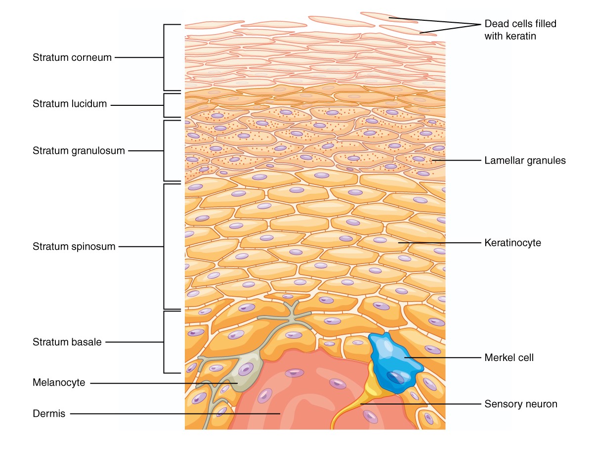
00:00 Well let's now look at skin appendages. 00:04 Again they are epidermal in origin. During development, the epidermis burrows down into the underlying dermis and becomes specialized into various structures. Shown here in this diagram is a picture of three-dimensional structure of skin. And it shows you on the right hand side diagram, the epidermis and hair projecting from the surface. 00:39 Underneath there are various other structures that are derived from the epidermis. Sweat glands of two types, eccrine and apocrine are also derived from the epidermis. They are examples of these epidermal appendages. We will talk about these in more detail in a moment. 01:01 Hair follicles that are areas where the hair is produced and renewed and associated with hair follicles are sebaceous glands. They are all skin appendages that have very important roles in the function of skin. 01:21 Let us look at the sweat gland in more detail. There are two types. Eccrine sweat glands that are widely distributed over the body and also the apocrine sweat glands that are restricted mainly to the locations of the body I've listed there, particularly the axilla. 01:40 Here is an eccrine sweat gland. It consists of a secretory portion that I have labeled here, a paler stained portion of the gland and a duct portion which is a darker stained area. Much smaller profile the duct portion has than the large, wider lumen secretory portion. If you look very carefully at the secretory portion, it is also surrounded by myoepithelial cells, which are contractile cells and they help to dispel the sweat. 02:24 On the right hand side image, you can see if you look towards the left hand edge of this image, the duct proceeding its way through the dermis and out through the epidermis. 02:38 Now the best way to imagine what a sweat gland looks like, I think you just think of a cooked piece of spaghetti. Next time you go home and cook spaghetti, get a piece of this spaghetti and hold it up on your hand and stand next to the edge of your kitchen bench and allow that piece of spaghetti until it comes in contact with the bench and then keep lowering it some more. What happens is the spaghetti will start to coil up. What you hold in your hand is the duct portion and what is coiled down the bottom on your kitchen bench is the secretory portion. So when you cut through that piece of spaghetti at various levels, you are going to see different profiles of the spaghetti. Such is what you see here in this image, you can see parts of the duct system cut transversely perhaps, sometimes longitudinally or obliquely, the same with the excretory portion. Now when we secrete sweat, we are doing two things. We are secreting water, which evaporates from the surface above skin. So sweat has a thermoregulatory role, that cools the body. The evaporation of that water or sweat from the surface cools the body. So one important function of sweat is thermoregulation. Other components of sweat are components of the body that we want to get rid of such are uric acid, urea, ammonia. And therefore sweat glands are also not just a thermoregulatory organ, but an excretory organ. Here is an apocrine sweat gland. 04:36 It has got a wider lumen, a large luminal space lined by cuboidal like epithelial cells. There are also myoepithelial cells wrapping around these secretory components that help to eliminate the secretion product. The ducts are usually the same size as what you see in the sweat gland but remember they do not modify the secretion product. Accrine sweat glands the ducts do modify the product. These apocrine sweat glands become active during puberty and they secretory proteins and pheromones. And these are odourless, but they can be acted upon by bacteria and create an acute odour. It is important to understand something about the innervation of these sweat glands. They are both innervated by sympathetic divisions of the autonomic nervous system. But in both situations, the neurotransmitter substance used is different. The eccrine sweat glands are cholinergic. They are said to be cholinergic whereas apocrine sweat glands are said to be controlled by adrenergic nerve fibres. And I will explain that in a lot more detail when I lecture on the nervous tissues. And that brings about different reactions because when we secrete sweat with the role of thermoregulation, we first of all sweat from our forehead, and then our scalp, and then the rest of our body and finally the palms of our hands and the soles of our feet. But when we sweat because we are emotionally activated, or we're stress, we start secreting sweat from both the eccrine sweat glands and also the apocrine sweat glands, particularly the axilla. There we tends to sweat first on the palms of our hands and the soles of our feet. So the difference stimuli for sweating whether it be by the eccrine sweat glands or by the apocrine sweat glands are controlled by separate types of fibres from the sympathetic nervous system, cholinergic and also adrenergic. Let us briefly look at the structure of hair and the hair follicle. It is not something that we emphasize a lot in histology of the skin, but shown here are three profiles through sections of the hair at different levels as they travel up through the dermis before the hair finally protrudes from the surface of skin. On the left hand side, you can see the dermal papilla, projecting into the base of the hair follicle. That dermal papilla is no different to the dermal papilla projecting up into the overlying layer of epidermis that I've described before. The very surface of that dermal papillae has on it epidermal cells, typical of the epidermis, stratum basale and also melanocytes. And they're going to contribute to the colour of hair. And then on the outside of the hair follicle and on the outside of the hair as it proceeds to the surface, are a number of root sheaths, the internal and the outer root sheath. The outer root sheath is really just a representation of the down growth of epidermis. And the internal root sheath is really a few layers of soft keratin, as it's produce and gives rise to the more firmer shaft of keratin in the center of the hair. And on the remaining two slides, the one in the middle and the one on the outside right hand side, you see a transverse section taken through the hair, showing different layers of the hair. On the far right-hand side, you see a central white unstained region, which represents the hair shaft and two layers on the outside that represent the hair cortical and the hair cuticle regions of the hair. 09:36 But again as I said earlier, we do not emphasize really a lot about the histological structure of hair in our lecture on skin. Associated with each of these hair follicles are sebaceous glands that I'd like to describe here. Sebaceous glands secrete sebum or oil. 10:02 It keeps out hair oily and it keeps the surface of our skin oily. And again these sebaceous glands are formed by downgrowths of epidermis, by downgrowths of the external root sheaths that I've mentioned earlier. And sebaceous glands are an example of holocrine secretion. 10:30 Holocrine secretion is where the cell accumulates its secretory product and then the cell dies. 10:38 It disintegrates by a process called programmed cell death or apoptosis. And here in this image, you can see the very basal parts of the sebaceous gland, the cells being derived from basal stem cells. They accumulate their secretory product, the sebum and then finally towards the luminal space associate with the hair follicle, the cells die and the oily secretory contents, the sebum is liberated into the luminal space.Let us finally look briefly at the nail. On the right-hand side, you see a section through the tip of the finger. You can see typical epidermis. On the top left hand part of this image, you can see a yellow stained component, that is the section through the nail, which is just keratin. Again formation of nail is just like the formation of keratin on the surface of our skin and also like the formation of the hair. But there's four or five major components of this fingernail or this section through the fingernail that I want to point out to you. First of all, the nail plate is a part of the skin where the epidermis loses its layer of keratin as it comes around to start to form where the nail is going to exit. And then along the nail bed all you have really is remaining components of the epidermis, the stratum basale and the stratum spinosum because you are not producing typical keratin that you do on the surface of skin, you are producing the nail. And underneath the nail is the nail root. That is the area that germinates the layers of keratin, germinates the cells that are going to go through the process of keratinization ,forming the nail root and the nail matrix. And as more and more keratin is formed, the fingernail is going to start to proceed and protrude from the surface of the finger. There are two parts of the fingernail that are very very important. That is the eponychium and the hyponychium. And those two components, the eponychium and the hyponychium are areas that seal the fingernail, the underneath surface of the fingernail from the exterior. 13:44 The eponychium is the cuticle region of the fingernail. It stops pathogens from getting into underlying germinal layers of the fingernail. And the hyponychium is where the nail protrudes from the finger. And immediately underneath that nail again is a region that you do not want pathogens to be able to access, the germinating layers of the nail bed and the nail root. 14:17 Well, I hope you have learned now a fair bit about the structure of skin. Make sure you understand the layers of the epidermis and all their functional significances. 14:29 Make sure you know the difference between the thick and thin skin and know all the functions of skin, particularly the functions that are served by all the cells that make up the epidermis, and the function of all the skin appendages. And also know how to recognize those cells and also those skin appendages. So thank you very much for listening to this lecture. Again I hope you have enjoyed learning about the histology of skin.
About the Lecture
The lecture Skin Appendages by Geoffrey Meyer, PhD is from the course Epithelial Tissue.
Included Quiz Questions
Which of the following statements best describes eccrine sweat glands?
- They are innervated by the sympathetic nervous system.
- Their highest density is on the skin of the trunk.
- They primarily respond to temperature changes in the palms and soles.
- They are associated with hair follicles.
- They have myoepithelial cells surrounding their duct portions.
What is the function of sweat glands?
- All of the other answer options are functions of sweat glands.
- Thermoregulation
- Excretion of electrolytes
- Protection of skin
- Excretion of water
The secretory portion of an apocrine gland is a coiled, nonbranching tube that is most commonly lined by which of the following?
- A layer of cuboidal cells
- Multiple layers of columnar cells
- A layer of squamous cells
- Multiple layers of transitional cells
- A layer of goblet cells
Which of the following constitutes the primary type of innervation in eccrine sweat glands?
- Cholinergic fibers of the sympathetic nervous system
- Adrenergic fibers of the sympathetic nervous system
- Cholinergic fibers of the parasympathetic nervous system
- Adrenergic fibers of the parasympathetic nervous system
Which of the following glands is a holocrine gland?
- Sebaceous gland
- Apocrine gland
- Eccrine gland
- Merocrine gland
- Mammary gland
Author of lecture Skin Appendages

Geoffrey Meyer, PhD
Customer reviews
5,0 of 5 stars
| 5 Stars |
|
1 |
| 4 Stars |
|
0 |
| 3 Stars |
|
0 |
| 2 Stars |
|
0 |
| 1 Star |
|
0 |
This is by far the best histology lecture there is online. love the way make it so easy to comprehend and the Q&A Are amazing
