Playlist
Show Playlist
Hide Playlist
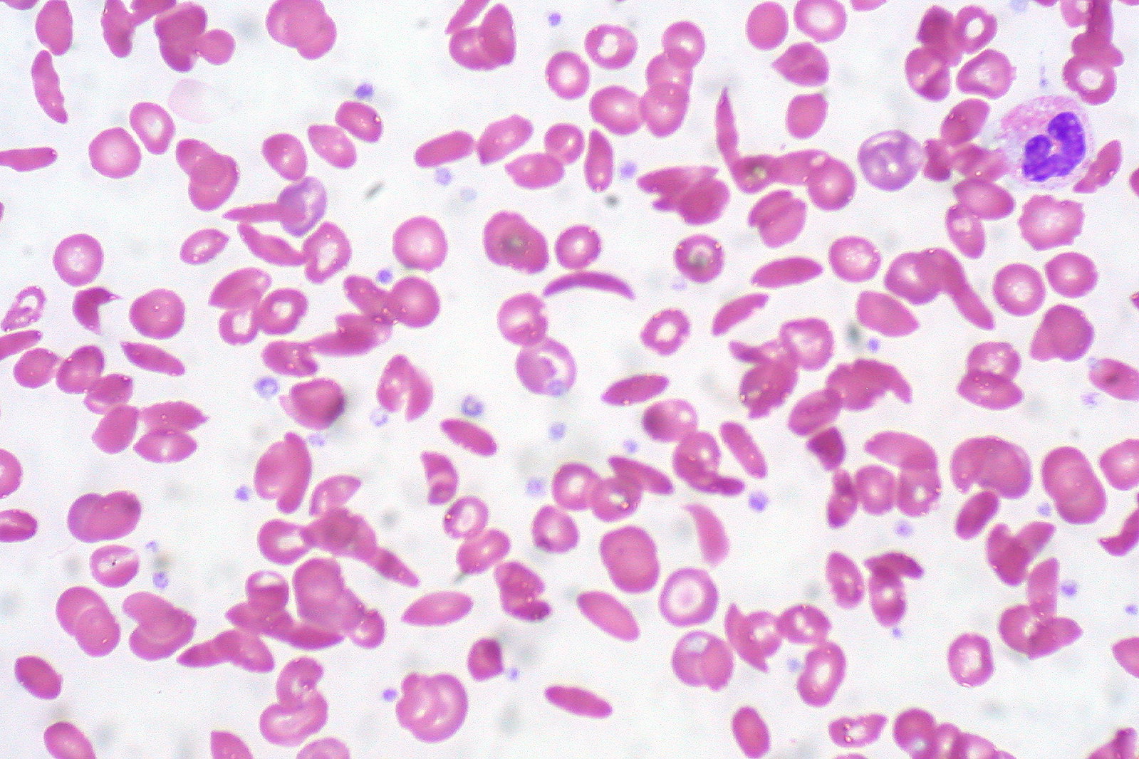
Sickle Cell Anemia: Etiology


-
Slides Sickle Cell.pdf
-
Reference List Pathology.pdf
-
Download Lecture Overview
00:01 Let’s take a look at the all important sickle cell anemia and how your patient is going to present. 00:08 To begin with, let’s get straight into the detail. 00:11 I want you to take a look at what’s bold in here. 00:14 You’ll find glutamic acid which is then being replaced by valine at position 6. 00:23 That will be the most important substitution that you want to be responsible for. 00:27 And in fact, you actually screen for this on your beta globin and you call this E6V. 00:33 And what E6V means, meaning to say like -- Nowadays, you can actually genetically screen for a patient that you would suspect as having sickle cell anemia by doing a glutamic acid E6 position for valine. 00:50 This gives HbS. 00:53 For every HbS that you then acquire then you end up having either a heterozygous or a homozygous type of pattern. 01:03 Obviously, whenever you have a homozygous type of disease, that is far more worse than a heterozygous. 01:09 Are we clear? It’s an autosomal recessive inheritance which means that you can only pick up one. 01:15 If you pick up one, you end up having what? Sickle cell trait. 01:19 That’s heterozygous. 01:21 If you pick up both, if homozygous then that will be sickle cell disease. 01:26 Make sure that you are quite familiar with the terminology and the language. 01:30 What you’re seeing here at the bottom, ladies and gentlemen, is a very important amino acid and a test in which the first one, on your left, is showing you hemoglobin A on your beta globin. 01:41 And you notice that the 6 position, well, there is my glutamic acid. 01:46 Do you see the 6 position in red? I would also know GAG from genetics. 01:52 The one on the right, you find that the glutamic acid is being replaced by valine. 01:57 At what position? Six. 01:59 This is GTG. 02:01 So the A is being replaced by T, the glutamic acid replaced by valine at the 6 position. 02:08 What did you form in the process? On the right, on the beta globin, take a look, you formed your hemoglobin S. 02:16 Move on.
About the Lecture
The lecture Sickle Cell Anemia: Etiology by Carlo Raj, MD is from the course Hemolytic Anemia – Red Blood Cell Pathology (RBC).
Included Quiz Questions
A patient with sickle cell anemia has which of the following point mutations?
- Glutamic acid replaced by valine at position 6 of the beta chain
- Glutamic acid replaced by valine at position 6 of the alpha chain
- Glutamic acid replaced by valine at position 21 of the beta chain
- Valine replaced by glutamic acid at position 6 of the beta chain
- Glutamic acid replaced by valine at position 21 of the alpha chain
Author of lecture Sickle Cell Anemia: Etiology

Carlo Raj, MD
Customer reviews
5,0 of 5 stars
| 5 Stars |
|
5 |
| 4 Stars |
|
0 |
| 3 Stars |
|
0 |
| 2 Stars |
|
0 |
| 1 Star |
|
0 |
