Playlist
Show Playlist
Hide Playlist
Short Muscles and Extrinsic Tendons – Anatomy of the Hand
-
Slides 07 UpperLimbAnatomy Pickering.pdf
-
Download Lecture Overview
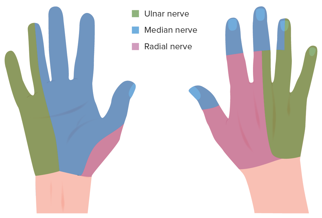
00:00 So now let's move on to the short muscles and how that arranged alongside the extrinsic tendons. So here we can see some deep dissections of the hand. We can see that we've cut away some of the tendons and we've opened up the carpal tunnel and here we have removed some of those tendons as well to look deep and see some dorsal and palmar interossei muscles. So if we have a look, we can see the all 8 tendons, see them here, flexor digitorum profundus. And we can see the cut tendons here, flexor digitorum superficialis, and to the central compartment of the hand by passing through the carpal tunnel. And if you remember, FDS, flexor digotorum superficialis, pass to the middle phalanx of digits 2-5. Whereas FDP, flexor digitorum profundus, pass to the distal phalanx of digits 2-5. So there's an important arrangement here that we need to observe. As the tendons or flexor digitorum superficialis pass to the middle phalanx, the tendons actually split and we can see that here. Here, we can see the tendon of FDS passing towards the middle phalanx and its tendons splitting. See it here? We can also see it happening here. See the tendons splitting into 2 as they go in to touch to the middle phalanx. We can see again here once these tendons are actually being reflected somewhat. So the tendon of flex digitorum superficialis here and we can see this actually splits, one going down onto this lateral side and one just going down this medial side. It's a little bit covered by these, the tendon of flexor digitorum profundus. The tendon of flexor digitorum profundus as we can see passes deep through this split. So as the tendon of flexor digitorum superficialis split, so the deeper tendon of flexor digitorum profundus pass through this split as it pass to the distal phalanx. And this is a really important arrangement. 02:18 We can see again here, flexor digitorum profundus is passing deep to flexor digitorum superficialis. And then as it splits, the tendon here is splitting FDP passes deep to it. 02:32 And this enables the tendon of FDP to pass to the distal phalanx. We also have some vincula tendinum. We have two of these and these are very small little slips of tissue and they actually attach the tendons of FDP and FDS to each other and also to the phalanx. We can see these small little tendinous slips. We can see them passing down here and here we have a longum and a brevia. We have a long one and we have a short one and these are important in connecting those 2 tendons to the tendinous sheaths and also in permitting some microcirculation. So these carry some micro blood vessels to the tendons for that blood supply. So they're really important in connecting the tendons to each other and also to the phalanges and also permitting some microcirculation so the tendons can receive that oxygen and nutrients. So these muscles are capable of working across 2 different joints. 03:43 We have a lumbrical here. We have another lumbrical here, are 2 lateral unipennate lumbricals. And then we have 2 bipennate lumbricals. We can see we have 1 here and we have 1 here. So we have 4 lumbricals together. Notice how they all originate from the tendon of flexor digitorum profundus and they don't work on the thumb, they don't work on the first digits or any associated with digits 2, 3, 4, and 5. So if we have a look, we can see lumbricals 1 and 2. These originating from the lateral 2 tendons of FDP and these are unipennate ones, I indicate it. And then we have lumbricals 3 and 4 and they're originating from the medial 2 tendons of FDP and these are bipennate. They insert onto the lateral surface of the extensor expansions, as I mention, of the digits 2 and 5. So they don't work on the thumb, just digits 2-5. The lumbricals 1 and 2, the lateral 2 lumbricals, are innovated by the median nerve whereas the lumbricals 3 and 4 associate with the medial tendons of FDP are innovated by the ulnar nerve. So we can see the lumbricals have a different innovation, a different nerve supply. The function of these lumbricals is important because they can both flex and extend different joints. So they can flex the metacarpophalangeal joint, the joint between the phalanges and the metacarpals, so they can flex that joint. The phalanges here and the metacarpals, they can flex that joint. And they are also capable of extending the interphalangeal joints. So they're capable of flexing that joint but also extending the interphalangeal joint. And that is because of their position. They run anterior to the metacarpophalangeal joint so they can flex it and by passing to the extensor expansion, they're running posterior to the interphalangeal joints. So contraction of these muscles allows flexion of that joint, the metacarpophalangeal and extension of the interphalangeal joint. And these position here is important if you're holding a pencil if you're about to write. So these muscles are important for the digits to assume complex positions. Now let's carry on looking at a series of short muscles, and these are known as your interossei muscles. 06:22 We have 2 different types of interossei muscles. We have dorsal interossei positioned on the dorsal aspect of the hand and we have palmar interossei muscles positioned on the palmar surface. Here, we can see the dorsal interosseous muscles. We have 4 of them. We can see 1, 2, 3, 4 and we can see these have 2 heads. They're running from the adjacent surfaces of all of the metacarpals. So we can see the dorsal interosseous here is running from the medial surface of the 1st metacarpal and the lateral surface of the metacarpal here at the 2nd metacarpal. And we have similar arrangements. And these are passing towards the extensor expansion, those extensor hoods over the digits. And then we have the palmar interossei. These are just running from one of the metacarpal surfaces and we can see they're coming from digits 2, they're coming from digits 4, and coming from digit 5. Digit 3 does not have an attachment of these interossei muscles. The interossei muscles do not attach. So we can see that these muscles are going to be associated with abducting and adducting the fingers. We can see the dorsal interossei are coming from the dorsal sides of all the metacarpals, these are bipennate muscles. And the palmar interossei, as I said, come from the palmar sides of metacarpals 2, 4, and 5. 08:02 They insert on to the base of the proximal phalanges and also the extensor expansions. The nerve supply for these interossei is by the ulnar nerve, the deep branch. And the dorsal interossei are associated with abducting digits 2-4. Palmar interossei are associated with adducting digits 2, 4, and 5 this time towards the axial line. I've put that here towards the axial line and away from the axial line. What does that mean? Well, if you imagine the axial line is running down in line with the middle finger, so an axial line is running down here. This is the line at which these fingers are going to be abducted or adducted. And we can see that with contraction of these muscles we can put the muscles in cartoon form here. With this axial line, we can see that the dorsal interossei are going to pull the fingers away from this axial line. So these dorsal interossei can pull away. This dorsal interossei can pull this middle finger away. It can also, because we have the dorsal interossei on the other side, abduct it the other way. For the fingers, we don't talk about abduction and adduction as moving them towards the midline of the body. We talk about moving away from this axial line. Therefore, the middle finger can both abduct this way and it can abduct that way. Whereas the other fingers will all abduct away from this axial line like that. The middle finger can abduct away either side. If we look at the palmar interossei, then we look at adducting, so palmar interossei adducting, and they are going to draw the fingers towards the middle finger, towards the axial line. So we can see that this interossei will move across, this interossei will move across, this interossei will also move across. So we've got our middle finger. We can adduct here, we can adduct here, and we can adduct here. So all the fingers are together. Don't forget we have adductor pollicis so we can move the thumb across. 10:18 It doesn't need an interosseous muscle. So here we can see the origins and insertions of these muscles. The easy way to remember the function is for the dorsal interossei to use the D from dorsal and the AB, so you can have DAB, dorsal interossei abduct. 10:36 And you can have PAD, using the P from palmar interossei pad as the palmar interossei adduct. So in this lecture, we've looked to the dorsal aspect of the hand, we've looked at the extrinsic extensor tendons and tendinous sheath. We've looked at extensor expansions and the anatomical snuffbox, its boundaries and contents. 10:58 We then looked at the palmar aspect, we looked to the carpal tunnel and the ulna canal. The boundaries and contents. We then looked at compartments, central hypothenar, thenar, adductor, and interosseous. And we looked to the muscles within each of the compartments. And then we looked to the extrinsic flexor tendons.
About the Lecture
The lecture Short Muscles and Extrinsic Tendons – Anatomy of the Hand by James Pickering, PhD is from the course Upper Limb Anatomy [Archive].
Included Quiz Questions
Which hypothenar muscle is located most medially within the hand?
- Abductor digiti minimi
- Abductor pollicis brevis
- Flexor digiti minimi
- Flexor pollicis brevis
- Opponens digiti minimi
Which statement concerning the lumbricals is correct?
- They flex the metacarpophalangeal joints and extend the interphalangeal joints.
- They extend both the metacarpophalangeal and interphalangeal joints.
- They flex both the metacarpophalangeal and interphalangeal joints.
- They extend the metacarpophalangeal joints and flex the interphalangeal joints.
- They only flex the metacarpophalangeal joints.
Which muscle is innervated by the ulnar nerve?
- Abductor digiti minimi
- Opponens pollicis
- Extensor digiti minimi
- 1st and 3rd lumbricals
- 1st and 4th lumbricals
Which muscle is found in the thenar eminence?
- Flexor pollicis brevis
- Second lumbrical
- Palmaris brevis
- Flexor pollicis longus
- Extensor pollicis longus
Which muscle has the tendons that are the origin of the lumbricals?
- Flexor digitorum profundus
- Flexor digitorum superficialis
- Palmaris longus
- Flexor digitorum indicis
- Flexor pollicis longus
Which statement regarding the lumbricals is correct?
- Lumbricals 1 and 2 originate from the lateral 2 tendons of the flexor digitorum profundus.
- Lumbricals 1 and 2 are bipennate.
- Lumbricals 3 and 4 are unipennate.
- Lumbricals 3 and 4 originate from the lateral 2 tendons of the flexor digitorum profundus.
- All lumbricals are unipennate.
Which nerve innervates the interossei muscles?
- Deep branch of the ulnar nerve
- Palmar cutaneous branch of the ulnar nerve
- Recurrent branch of the median nerve
- Superficial branch of the ulnar nerve
- Median nerve
Which digit is free from the palmar interossei muscles?
- 3
- 5
- 2
- 4
- 1
Customer reviews
5,0 of 5 stars
| 5 Stars |
|
1 |
| 4 Stars |
|
0 |
| 3 Stars |
|
0 |
| 2 Stars |
|
0 |
| 1 Star |
|
0 |
Good clear explanation of all the small muscles in the hand.


