Playlist
Show Playlist
Hide Playlist
Secondary Dilated Cardiomyopathy – Cardiomyopathy

-
Slides 05 Cardiology Alpert.pdf
-
Reference List Cardiology.pdf
-
Download Lecture Overview
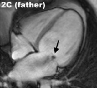
00:01 Ischemic cardiomyopathy, along with hypertrophic cardiomyopathy, are the two commonest forms of cardiomyopathy seen. In the dilated cardiomyopathy due to ischemic heart disease, very often the patients will have had symptomatic myocardial infarcts, usually multiples, and each one of these damages the left ventricle with the end result that one ends up with markedly reduced left ventricular muscle and a dilated cardiomyopathy. Something that we don’t understand is that some patients show up for the first time with heart failure, never having had a clinical heart attack or myocardial infarct, never having had angina, and yet when we do their coronary angiogram as part of the evaluation, we discover terrible, severe, advanced coronary disease as the cause. Why these patients have no symptoms and other patients have clear-cut symptoms is still a mystery and is the subject of intense investigation. 00:52 Hypertension, untreated, can also lead to dilated cardiomyopathy by putting a major stress on the left ventricle that eventually causes it to fail and dilate. Valvular heart disease, longstanding mitral regurgitation, longstanding aortic regurgitation, untreated, also eventually can lead to a dilated cardiomyopathy. The next one listed on your list here, the anthracyclines, these are drugs used in a variety of cancers, but particularly for breast cancer, particularly metastatic breast cancer, and they can cause heart damage. And so, the cardiologists are often involved with the oncologist in carefully monitoring the patient who’s receiving these drugs and we stop the treatment as soon as we start to see some heart injury. There’s a small number of women who, following pregnancy, will develop a dilated cardiomyopathy. Again, there seems to be a genetic predisposition here. And finally, chronic, heavy use of alcohol leads to a dilated cardiomyopathy, among other problems. 01:57 Now, let’s talk a little bit about ischemic dilated cardiomyopathy because again, this is a very very common form. It seems that this is the most common form of dilated cardiomyopathy, although the genetic forms now compete with it for most common status. It’s, as we’ve talked about, caused by repeated injuries from ischemia that can be symptomatic or asymptomatic. And, in fact, many of these patients come first to attention when they have a myocardial infarction that is symptomatic. But others, as I’ve told you, come to attention when they’ve never had any symptoms and we discover, to our surprise, that the patient has severe coronary artery disease. The prognosis is not good with the dilated cardiomyopathy, as I’ve been expressing throughout this section. 02:47 Any form of dilated cardiomyopathy has a poor prognosis. The ischemic form has the worst prognosis. Now, let’s talk for a few moments about diagnosis. We would, of course, love to diagnose these patients early in the course of their cardiomyopathy. Unfortunately, they usually come to our attention when they develop symptoms of quite significant heart failure. And, of course, in the heart failure segment, we’ve already talked about that. Patients may be short of breath. They may have peripheral swelling and edema. Sometimes the ischemic form, the patient will have had some ischemic event, and then when we do the echocardiogram and the catheterization, we discover, to our surprise, that the left ventricle has been severely injured, has a markedly reduced ejection fraction that is a reduced percentage of blood that it squeezes out with each beat. There are often electrical abnormalities, both atrial and ventricular arrhythmias. We’re going to talk about the arrhythmias in a few moments. And, of course, there are enla-, there’s enlargement on the electrocardiogram of the left atrium and also, often evidence for enlargement of the left ventricle as well. On physical exam, it’s the heart failure exam. The heart may feel enlarged when you feel the chest. There is often a third heart sound which is a sign that the ventricle has been severely damaged. There may be sounds in the lung that suggest fluid. There may be edema in the legs, as I’ve already said. 04:25 And one may even have, as I mentioned before, mitral or tricuspid regurgitation murmurs from stretching of the valve annulus or ring in which the valve is contained. The workup for the diagnosis usually starts, of course, with the physical exam and a history, followed by an electrocardiogram. Usually, by then, we’re starting to have a clue because we see it's a heart failure patient, that we’re dealing with a dilated cardiomyopathy. 04:52 And then the echocardiogram shows us the picture that really confirms it. We usually do a coronary angiogram to distinguish the ischemic form from the forms that are not ischemic, for example, the genetic or alcoholic ones or other etiologies. Occasionally, we’ll do a magnetic resonance image because it gives us an idea about what is the underlying structure of the myocardium. There might be a clue, “Oh, this is ischemic,” or “No this is secondary to a chemical that’s being used for treatment of cancer,” or could be from alcohol. These days, we’re doing more MRIs because they give us a lot of information about the structure of the myocardium in patients with dilated cardiomyopathy. And again, coronary angiography to make sure we’re not dealing with ischemic cardiomyopathy. In some, not very many, individuals with dilated cardiomyopathy, when we do coronary bypass and and give new blood flow to the heart muscle, some of the heart muscle recovers. And so, there’s partial recovery of the ischemic cardiomyopathic patient, but usually, that doesn’t happen. Usually, the ventricle is too damaged and scarred to recover and after medical therapy fails to work, these patients are often candidates for heart transplant. But, we’ll talk about that in a few minutes. So, the therapy, of course, is the same therapy we talked about for heart failure. 06:21 Our treatment strategy has three main components. 06:25 Medical therapy, advanced options for selected patients and device therapy. 06:29 For selected patients for medical therapy. 06:31 We start with beta blockers, which are crucial as they reduce both overall mortality and sudden cardiac death risk for the renin angiotensin system. 06:41 Our preferred agent now is sacubitril valsartan, what we call an Arni. 06:46 This has shown superior outcomes compared to traditional Ace inhibitors or ARBs, though these remain important options when Arni isn't suitable. 06:56 A major advance has been the addition of Sglt2 inhibitors to our armamentarium. 07:01 These medications have shown remarkable benefits in reducing both heart failure, hospitalizations and mortality. 07:08 Even in non-diabetic patients. 07:11 For mineralocorticoid receptor antagonists MRAs, we have both traditional options like spironolactone, (Aldactone) and and eplerenone and now a newer agent, finerenone. 07:25 All of these medications help decrease edema tendency in heart failure patients, though they work in slightly different ways. 07:32 The traditional agents have long proven benefits, while finerenone being nonsteroidal offers the advantage of less hyperkalemia and fewer hormonal side effects. 07:43 The choice of which MRA to use often depends on individual patient factors like kidney function and tolerance. 07:52 Of course, beyond medications, we emphasize the importance of reducing salt in their diet, and we often find that regular exercise helps when appropriate. 08:02 We always have to pay careful attention to electrical abnormalities arrhythmias. 08:07 This is where device therapy comes in. 08:10 Cardiac resynchronization therapy, or CRT, plays a vital role for specific patients. 08:18 When we see a wide QRS complex on ECG, typically 150 milliseconds or greater, implanting a special type of pacemaker can help re coordinate the left ventricular contraction, often improving heart function significantly. 08:34 Finally, for patients who continue to deteriorate despite optimal medical and device therapy, we have advanced options. 08:42 These include left ventricular assist devices, artificial hearts, and ultimately cardiac transplantation. 08:50 However, these are reserved for carefully selected patients. 08:54 They need to be sick enough to require such aggressive intervention, but healthy enough to benefit from it. 09:00 The key to success is a systematic stepwise approach, starting with optimal medical therapy and lifestyle modifications. 09:08 Adding device therapy when appropriate and moving to advanced options only when necessary. 09:14 Each step builds on the previous ones, and we need to ensure we've optimized each level before moving to the next.
About the Lecture
The lecture Secondary Dilated Cardiomyopathy – Cardiomyopathy by Joseph Alpert, MD is from the course Cardiac Diseases.
Included Quiz Questions
Which of the following findings is less likely in a patient with ischemic dilated cardiomyopathy without significant obstructive valvulopathy?
- High-pitched murmur in the base of the heart radiating to the carotids
- A narrow pulse pressure
- 3ʳᵈ heart sound
- Enlarged and displaced apex beat
- 4th heart sound
Which of the following drugs is most likely to cause dilated cardiomyopathy at therapeutic levels?
- Doxorubicin
- Propranolol
- Levothyroxine
- Prednisone
Which of the following is not a treatment option for dilated cardiomyopathy?
- Pericardiectomy
- Ventricular assist device
- Cardiac transplantation
- Angiotensin-converting enzyme inhibitors
- Furosemide
Author of lecture Secondary Dilated Cardiomyopathy – Cardiomyopathy

Joseph Alpert, MD
Customer reviews
3,6 of 5 stars
| 5 Stars |
|
3 |
| 4 Stars |
|
0 |
| 3 Stars |
|
0 |
| 2 Stars |
|
1 |
| 1 Star |
|
1 |
Very good concepts are explained in these lectures. Classification is explained well in the first lecture with a flow diagram.
The topic it's well explained, clear and highly related to previous lectures.
mixed information,make me lost.i need to read another reference to understand this confusion
Good lecture. dont understand why someone gave it only a one
