Playlist
Show Playlist
Hide Playlist
Sebaceous Gland Tumors: Pathophysiology

-
Reference List Pathology.pdf
-
Slides Sebaceous Gland Tumors Pathophysiology.pdf
-
Download Lecture Overview
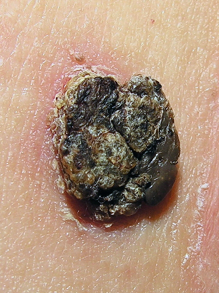
00:01 Welcome. In this talk, we will start delving into some of the malignant lesions of the skin. 00:09 Some of these are quite common, some of them not very common at all. 00:13 This group, specifically the sebaceous gland tumors, are not all that common, but we will talk in this one both about benign lesions benign tumors and malignant tumors. 00:26 So sebaceous gland tumors are very rare neoplasms and they derive from the sebaceous glands. The pictures you see are indicative of a couple different variants. But all of them need a diagnosis by pathology in order to ascertain exactly what is going on. 00:44 All right, just by way of a quick review, remember that sebaceous glands are glands that are attached to the hair shaft, the hair root. 00:54 And in fact, in that location is where we will have a variety of of skin stem cells as well melanocyte stem cells and epidermal stem cells. The sebaceous glands attach to the hair shaft and hair root are going to secrete their products as a holocrine secretion containing fat, sebum, variety of proteins onto the surface of the hair shaft and onto the surface of the skin, and that provides an emollient kind of surface. 01:25 It also limits the loss of water. 01:28 So that's what the sebaceous gland does. 01:30 And the histologic image demonstrates areas where the sebaceous glands are. 01:36 So they are way down along the hair shaft and we find them predominantly within the dermis. 01:42 Okay. Sebaceous gland tumors benign and malignant. 01:46 The benign versions are just vary from a hyperplasia just an increased proliferation to an actual discrete tumor that's still benign. It doesn't have the features of malignancy. It doesn't have genetic instability. So we would call those sebaceous adenomas. 02:02 Or if you're being really erudite, sebaceoma. 02:06 The malignant version are sebaceous carcinomas. 02:09 All right. The epidemiology of these overall they are pretty rare. Sebaceous hyperplasia can be seen in 1% of the healthy population. But in most cases we wouldn't even even know that that was happening. 02:23 To be perfectly honest, Sebaceoma has occurred more frequently in women than in men, suggesting that there may be an endocrine component in terms of the proliferation of sebaceous cells. 02:37 And they typically occur in an older population, so reflecting perhaps more of a lifetime exposure to hormonal stimulation. 02:45 Sebaceous carcinomas, on the other hand, are pretty infrequent. 02:48 They range between a very low number to maybe 5% of all malignant cutaneous tumors. 02:55 I think that that depends on the different populations that you're looking at. 02:59 However, if you're looking at tumors of the eyelid, then it is a reasonably common cause of eyelid malignancy, and patients tend to be older adults. 03:10 The pathophysiology of this. 03:12 So for hyperplasia or the sebaceoma they can be they can occur sporadically due to UV exposure. And then with the perhaps spontaneous acquired mutation of genes that are going to regulate proliferation within the sebaceous glands. Keep in mind, sebaceous glands are actually fairly deep within the dermis, so it's hard to get a whole lot of UV exposure to them. 03:36 Nevertheless, it does occur. 03:38 You can also have congenital sebaceous gland hyperplasia or sebaceomas , in the setting of Muir-torre syndrome. 03:45 And that's basically a variant on Nonpolyposis syndrome, the Lynch syndrome, which is a variety of mutations in mismatch repair genes. Other inactivating mutations in some transcription factors or in some tumor suppressors can also drive this, including the lymphoid enhancer transcription factor one. 04:07 Given the rarity of this overall, I think that it's enough to remember that it can occur sporadically and can be benign. 04:15 The malignant version have additional somatic mutations. 04:19 So these have acquired not only a driver mutation for proliferation, but additional mutations that are going to give rise to genetic instability and the acquisition of additional phenotypic changes that will make these tumors invasive and prone to metastasis. 04:38 So it can begin with microsatellite instability pattern those reflecting mismatch repair gene mutations. 04:44 But we can also have ultraviolet damage and certain mutations that are recognizable in notch or FAT3 or TP53, or any of a variety of genes may suggest a UV damage driving this. 05:02 Again, given the overall rarity of these tumors, I don't think it's important that you necessarily remember those genes. 05:08 You can also have very minimal mutations in some of the ocular those associated with the eyelids sebaceous carcinomas. 05:18 Clinical presentation. 05:19 So sebaceous hyperplasia often kind of just overlooked. 05:23 It's a ditzel that people don't really worry about too much. 05:26 They can be they tend to be rather small. 05:30 2 to 6mm. The center tends to be somewhat umbilicated with a little kind of dimple within it, they are skin colored, and therefore may not even be all that apparent. They do appear in a place where you might see them, such as the forehead, the glabella, the space between the eyebrows, and the nose, nose, and cheeks of older individuals. 05:49 Remember, it occurs more as you age. 05:52 Sebaceous adenomas and sebaceomas, so these are the benign greater benign tumors with more proliferation there. 06:01 They are still very well differentiated. 06:03 They haven't acquired additional mutations. 06:05 All they're doing is proliferating. 06:07 They tend to be located very much in the upper dermis, suggesting perhaps a UV causation. 06:14 They're solitary. They tend to be larger than hyperplasia, as you see here, your yellowish to brownish. 06:20 Again, if you think about it, what they're producing is a bunch of sebum. 06:24 That sebum can get oxidized. 06:25 So it can have kind of a brownish coloration. 06:27 And like the sebaceous hyperplasia tend to be located on the head and neck. Finally sebaceous carcinomas. 06:35 Those are the ones that you should be worried about when you see a lesion in the eyelid. 06:39 They're very they're relatively common when they do occur in the head and neck region, particularly around the eyes. 06:45 They tend to be painless. 06:47 They're rounded nodules, most typically in the upper eyelid. 06:51 Not clear why that is, except that maybe the upper eyelid gets more UV than the lower eyelid. 06:56 Don't know. They are yellowish and tannish nodules. 07:00 There may be some hyperpigmentation, but also oxidation of sebum that's there. 07:04 They're often ulcerated. 07:06 And that is a bad prognostic sign suggesting that it's not just hyperplasia or a sebaceoma, but in fact we've gone malignant. 07:15 The diagnosis is going to require a biopsy. 07:17 So a lot of other things can kind of look like these lesions grossly including on the eyelid. 07:23 So you want to do an excisional biopsy in some cases. 07:27 In areas like the eyelid you want to do a punch biopsy and then send it off for histopathologic evaluation. 07:33 What we're going to be seeing, it ranges all the way from just hyperplasia for that adenoma, which and everything is well differentiated and it's not going anywhere to now an invasive, highly atypical appearing lesion for sebaceous carcinomas. 07:52 And where it has expanded beyond the basement membrane, it is inducing an inflammatory and or fibrotic response. 07:59 And it is impinging on nerves and or lymphatics. 08:03 And those are all bad signs suggesting that we're probably dealing with a malignancy. If there are multiple of these, you may want to screen for the Muir-torre syndrome again. 08:14 That's a variation on the on the mismatch repair gene mutations. Regarding management. 08:22 So for benign lesions a simple surgical excision will do, especially if it's in an area, say between the eyebrows on the face that you really don't want to have a lesion. 08:33 In some cases where you just want to make the diagnosis and see whether it's benign or malignant, you may not have to take the whole thing off. 08:40 But a partial biopsy followed by clinical follow-up for those who don't want to have major or more surgery to remove the whole thing. 08:48 For malignant lesions, we need to reduce the size of them, initially they are somewhat radiation sensitive. 08:55 So we would do that. And then we would want to do a complete surgical excision. 08:59 With that we've dealt with a relatively uncommon but nevertheless important lesion sebaceous gland tumors both benign and malignant.
About the Lecture
The lecture Sebaceous Gland Tumors: Pathophysiology by Richard Mitchell, MD, PhD is from the course Adnexal Tumors of the Skin.
Included Quiz Questions
Where are sebaceous glands primarily located in relation to hair structures?
- Attached to the hair shaft within the dermis
- Above the epidermis on the skin surface
- Within the subcutaneous fat layer
- Between the epidermis and dermis
- Inside the hair follicle bulb only
Which best describes the typical presentation of sebaceous hyperplasia?
- Small 2-6mm umbilicated lesions on the forehead and nose
- Large ulcerated masses on the trunk
- Painful nodules on the extremities
- Pigmented patches on the scalp
- Fluid-filled vesicles on the neck
Which location is most commonly associated with sebaceous carcinoma?
- Upper eyelid
- Lower extremities
- Lower back
- Chest wall
- Palms and soles
Which underlying genetic abnormality is associated with sebaceous tumors in Muir-Torre syndrome?
- Mutations in mismatch repair genes
- BRAF gene mutations
- Keratin gene mutations
- Collagen gene mutations
- Melanin synthesis gene mutations
What is the preferred diagnostic approach for a suspected sebaceous tumor of the eyelid?
- Punch biopsy with histopathologic evaluation
- Clinical observation only
- Dermoscopy without biopsy
- Fine needle aspiration
- Imaging studies alone
Which feature most strongly suggests malignant transformation in a sebaceous tumor?
- Ulceration with invasion beyond the basement membrane
- Yellow-brown color
- Central umbilication
- Small size
- Dermal location
Author of lecture Sebaceous Gland Tumors: Pathophysiology

Richard Mitchell, MD, PhD
Customer reviews
5,0 of 5 stars
| 5 Stars |
|
5 |
| 4 Stars |
|
0 |
| 3 Stars |
|
0 |
| 2 Stars |
|
0 |
| 1 Star |
|
0 |
