Playlist
Show Playlist
Hide Playlist
Scrotal Imaging: Hydrocele and Varicocele

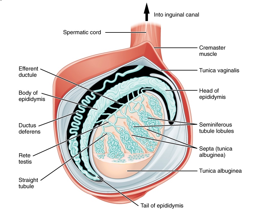
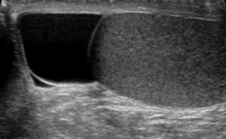
00:01 So in this lecture we will be discussing normal scrotal anatomy and abnormalities that are commonly seen within the scrotum. 00:07 The scrotum is best evaluated by ultrasound. 00:11 Usually scrotal ultrasound is performed for acute scrotal pain or any kind of palpable mass that the patient comes in with. 00:19 Common scrotal abnormalities include epididymitis and orchitis which is one of the most common things that are found, hydrocele, varicocele or enlargement of the vessel within the scrotum, testicular torsion and much less commonly testicular mass. 00:36 Let's go into each of these in a little bit more detail. 00:38 So this is an example of a normal scrotal ultrasound. 00:42 You can see that the testicle had this homogeneous echotexture with no focal lesions or surrounding fluid. 00:49 This right here is the measurement of the epididymis which is also very commonly seen on a normal scrotal ultrasound. 00:55 This is an example of normal Doppler waveform. 00:58 So whenever we're looking for blood flow to the testicle, we perform a Doppler ultrasound and this shows you arterial and venous flow to the testicle. 01:06 So the color images demonstrate the different variations of color flow within the testicle while the spectral images here demonstrate a normal venous spectral waveform which is approximately of equal height here, and a normal arterial waveform which is seen as a peak and then a down slope. This indicates that there is normal flow to the testicle. 01:25 So let's take a look at epididymitis and orchitis. 01:30 Here we have an image of a normal testicle. 01:32 You can see how the testicle is in relation to the epididymis. 01:37 Up here we have the epididymal head as well as the epididymal body and tail which kind of surrounds a portion of the testicle here. 01:44 And we can see multiple vessels flowing within it. 01:46 Epididymitis and orchitis is inflammation of the epididymis and or testis. 01:52 Either one can be inflamed. 01:53 So findings of epididymitis on imaging include an enlarged epididymis, increased blood flow to the epididymis as seen in Doppler ultrasound, a decreased echogenicity of the epididymis so it appears hypoechoic and this maybe focal or it may be diffused and orchitis is often seen secondary to the spread of infection from the epididymis so it may start off as an epididymitis and then if it is untreated, it can spread to the testicle and cause an orchitis as well. 02:23 So let's take a look at these ultrasound images. 02:26 What do we see here? So the first is an images of the testicle without any Doppler flow to it. 02:32 The second is an image with Doppler on and the third is just a different projection of the scrotum with Doppler on again. 02:40 So what do you see here? This is actually an example of epididymal orchitis. 02:51 We have an enlarged heterogeneous appearing epididymal head with increased blood flow. 02:56 The image of the testicle also demonstrates increased blood flow. 03:00 The entire testicle actually appears a little bit heterogeneous here. 03:03 And here we have significantly increased blood flow within the testicle. 03:07 So let's move on to hydrocele. 03:09 A hydrocele is fluid within the scrotum that surrounds the testicle. 03:13 It maybe congenital, but it may also be a result of other acquired abnormalities. 03:19 So it could be a result of epididymitis. 03:21 It could be also caused by torsion. 03:23 It can be caused by cancer within the testicle. 03:26 And it can also be caused by trauma. 03:28 And you wanna take a look at the type fluid that's in there. 03:31 You wanna take a look at whether the fluid has any internal echoes or whether it's simple appearing fluid and that can give you a hint as to what might be causing it. 03:38 Any of the abnormalities listed below maybe, may end up causing complex fluid or fluid with echoes within it. 03:45 So if it's simple fluid then it's more likely to be congenital. 03:47 So this is an example of simple appearing fluid surrounding the testicle. 03:52 So this is the normal scrotal sac and then here you see a large amount of fluid surrounding this testicle. 03:59 This fluid is simple appearing and you can see that the testicle otherwise appears normal. 04:05 So large hydrocele in a patient presenting with an enlarged scrotum. 04:09 This is often the complaining symptom of patients coming in, in patients that have hydroceles. 04:13 They have enlargement of the scrotum and they are making sure that there's no mass in there. 04:17 So varicoceles result from retrograde flow into the gonadal vein and that causes the veins of the pampiniform plexus to dilate to greater than 2 to 3 millimeters. 04:28 So normally we can see veins up to about 2 millimeters in diameter. 04:31 Anything greater than that and you're worried about the varicocele. 04:34 So you can see on this images here, this is the normal pampiniform plexus veins and these is when they become enlarged and we can see this on ultrasound. 04:43 So let's take a look. Varicoceles are best seen when you compare resting and valsalva images of the scrotum. 04:51 This is actually the most common cause of male infertility because it increases the heat to the testicle and that can cause infertility. 04:58 This is an example of a varicocele seen on ultrasound. 05:01 You can see here there's a measurement of the vein and it's actually dilated to over 3 millimeters so this indicates that there's a varicocele. 05:07 In this patient we have a comparison of Doppler resting and valsalva images and you can see here that there is some blood flow increased dilatation of the veins and some blood flow at rest but when you do valsalva, the blood flow significantly increases and this is a classic sign of a varicocele. 05:25 So valsalva again demonstrates increased distention of the vessels and that's because you have increased retrograde flow into the pampiniform plexus. 05:32 Testicular torsion is actually a do not miss finding. 05:38 You really wanna be able to find that immediately because detorsion can salvage the testicle and delaying this can actually lead to the loss of the testicle. 05:46 So some imaging finding of testicular torsion include overall decreased or absent blood flow to the testicle. 05:51 That's really the number one sign. 05:53 However, you can initially start off with just absent venous flow and then arterial flow is lost afterwards. 05:59 The testicle may appear enlarged and heterogeneous. 06:03 It can appear diffusely hypoechoic, and may also if you have a very hypoechoic testicle and it becomes small in size then that could actually indicate infarction. 06:13 In which case it would be too late to salvage the testicle.
About the Lecture
The lecture Scrotal Imaging: Hydrocele and Varicocele by Hetal Verma, MD is from the course Abdominal Radiology. It contains the following chapters:
- Scrotal Imaging
- Hydrocele
Included Quiz Questions
A patient presents with scrotal pain and an ultrasound shows increased blood flow to the epididymis and testicle with an enlarged epididymis. What is the diagnosis?
- Epididymo-orchitis
- Testicular cyst
- Testicular carcinoma
- Varicocele
- Testicular torsion
Which statement is FALSE regarding varicocele?
- Blood flow to the testicle is decreased or absent.
- It is due to the retrograde flow of blood in the gonadal vein.
- Pampiniform plexus dilates to greater than 2-3 mm.
- It is best seen with resting and Valsalva images of the scrotum.
- It is the most common cause of male infertility.
Which of the following is NOT seen with epididymitis?
- Decreased blood flow to the epididymis
- Enlarged epididymis
- Decreased echogenicity of the epididymis
- Orchitis often secondary due to the spread of infection
- May be focal or diffuse
Author of lecture Scrotal Imaging: Hydrocele and Varicocele

Hetal Verma, MD
Customer reviews
5,0 of 5 stars
| 5 Stars |
|
5 |
| 4 Stars |
|
0 |
| 3 Stars |
|
0 |
| 2 Stars |
|
0 |
| 1 Star |
|
0 |
