Playlist
Show Playlist
Hide Playlist
Retinoblastoma – Carcinogenesis

-
Slides Carcinogenesis Basic Principles.pdf
-
Download Lecture Overview
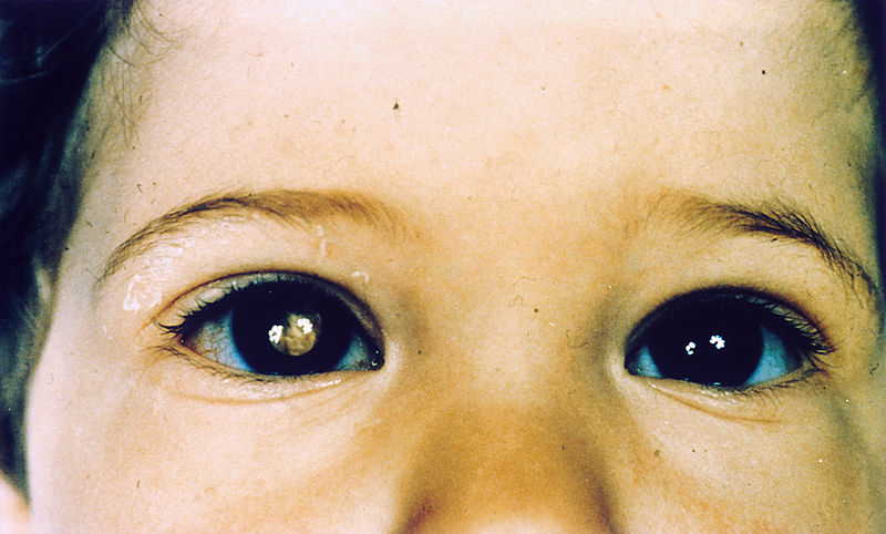
00:01 Now retinoblastoma let me show you a picture here so that you clearly see what I am referring to with Knudson two hit hypothesis. I want you to take a look at the patient on your left. This is the familial form. 00:13 I want you to focus upon that, there is a mother and you see the father. You see the Greek signs. 00:18 On your left is the father, on your right is the mama. The father here already has one hit. 00:26 I want you to focus upon the colours of the chromosomes. It seems as though that all chromosomes that are orange are normal. The blue represents the mutation. That is your one inherited hit. How many more hits do you require to develop cancer? One more. Go down the lineage. And you find, you see the bolt of lightning down there at the bottom, that one hit was all that was necessary to bring about a second, therefore, take a look at the retina in this patient. 00:59 That's retinoblastoma. Familial retinoblastoma. On the right, will be your sporadic form. Set up the picture again. 01:09 The father on your left, the mother on your right. You'll notice that all chromosomes are orange, all normal. Go down the lineage. 01:20 Progeny and you come to one lightning bolt, I'm just being silly here but that's your one hit. And then a sporadic second one comes along. Two hits now have taken place, sporadically without any inheritance. This is the sporadic form of retinoblastoma. 01:41 Sporadic, familial. I've given you two examples of familial thus far. They include familial retinoblastoma. 01:48 Here is familial. What I talked about p53, that's Li-Fraumeni. There is also others including familial melanoma. 01:58 We'll do special focusing on p53. p53. RAS was associated with 25,30,40% of cancers. p53 associated with 50% of all cancers. 02:12 Now keep in mind that you do not have to have just one mutation to bring about cancer majority of the time. 02:18 So maybe there is a mutation in RAS, maybe there is a mutation in p53, maybe there is a mutation elsewhere ultimately giving rise to lung, colon, breast. What I am saying is there will be many markers in which they will give you on your boards so that you know what kind of cancer you are going on to unless it's something like familial. The special cases. I did one for you already. 02:40 I did the one where the same patient had many familial cancers and that was Li-Fraumeni. In response to cell DNA damage, what is it going to do? It will do everything in it's power to make sure that the cell is properly repaired or worst case scenario, it will remove it out of the cell cycle permanently. Welcome to execution. Programmed cell death, aka apoptosis. Would you please tell me what the name of the enzyme is that then results in apoptosis. Caspase 9. I want us to do special focus on APC. Let's begin. 03:24 Where are you in the body. I want you to go into the intestine, that's where we are. So what are these? These are intestinal epithelial cells. These are enterocytes. If I tell you that this enterocytes are going to proliferate like crazy, what are you going to form? What are you going to see on lower endoscopy. What are you going to see please? A polyp. Okay. 03:49 Depending as to what kind of polyp you develop, you're increased in risk of, cancer. What kind? Colorectal cancer. 03:59 With all that said, now that you understand where we are, now let's take a look at the molecular mechanisms. 04:05 Our focus is APC so therefore focus on APC please. The first cell on your left is normal. No proliferation. By proliferation, what are you going to see again on scope. A polyp. And let's make this worst case scenario and we'll say villous. Or even worst case why not familial adenomatous polyposis. Because that's where we are headed. Familial adenomatous polyposis. FAP. 04:31 Normal cell on the left APC, it controls beta catenin. Take a look at the nucleus in the normal cell. TCF stand for transcription factor. And as long as APC is present, then beta catenin is controlled or perhaps even dissolved, therefore, there will not be excessive transcription. Next, I do want you to focus upon, the, well, be careful students get this confused. 04:59 I can tell you this. That this actually a derivative of a desmosome, okay. But where are you? In an enterocyte. 05:09 Watch this. You are going to love this. What's the name of that condition. We're going to knock out two birds with one stone. 05:15 What's the name of the condition where you take a tongue depressor and you scrape the skin and the skin comes right off. 05:21 It is a very vulgar disease. Pretty much gave it away. Pemphigus vulgaris. It's a type II hypersensitivity reaction. 05:29 IgG is attacking whom? The desmosome. So Dr. Raj is this a desmosome? No. Why, what's a desmosome? That's in your keratinocyte but attaches cell to cell. I'm telling you molecularly, E cadherin is a desmosome derivative. But what is it attaching? Not keratinocyte but enterocytes, epithelial cells. Is this clear? I'll come back to e cadherin. I don't want you to overlook it though. 06:01 Big time important for you. In the meantime though let's go on to develop familial adenomatous polyposis. Fun. 06:08 So here we have an APC mutation. You see that APC, the orange structure. It's gone away. Now beta catenin has been released, it's working in the nucleus to then bring about transcription. Worst case scenario, what if APC is gone, it's mutated, chromosome 5. If that APC has been mutated take a look at the last cell and APC has been completely mutated. Nothing regulating beta catenin. 06:38 How much transcription activity are you going to have? Relentless, endless. Therefore, how many polyps are you forming? Hundreds upon thousands. I'll show you a picture in gastroenterology where it's the 'carpet' of polyps. When you have a carpet full of polyps, what is your risk of colorectal cancer? 100%. Your order of hierachy of learning. APC, beta catenin and you have another receptor on the very top here. That receptor that is responsible for communicating with APC and beta catenin is called WNT. I don't care how you do it. Memorise it. If there is increase activity or relentless activity of WNT, there is going to be increased activity of your nucleus and therefore bringing about polyposis. I told you not to overlook e cadherin. That's the second time i'm bringing this to your attention. In neoplasia earlier when I was talking to you about, with metastasis and such I was talking to you about e cadherin. Your special focus in the word is adhere. Okay. 07:52 And that will adhere, your enterocytes normally together. Two major cancers right now that I want you to focus upon in which e cadherin is negative. First, the primary gastric adenocarcinoma that is spreading to the left supraclavicular lymph node. 08:09 Then it goes to the skin and it's called Leser-Trelet sign. And maybe hematogeneously will spread to the ovary. Krukenberg. 08:18 This is the diffuse type of gastric adenocarcinoma. Earlier, and I've mentioned this a few times. What's the number one prognostic indicator for breast cancer? Spread to the axillary lymph nodes. In lobular type of invasive breast cancer, e cadherin is usually negative, and you find that the breast cancer here loves to spread. You've lost your ability to adhere. Here is your full picture of APC. 08:45 Make sure that you know it in great detail. Let's move on. Patients with mutations in APC, it's chromosome 5. 08:53 At least know chromosome 5 develop FAP, familial adenomatous polyposis in which you would have relentless transcription activity and forming a carpet of polyps.
About the Lecture
The lecture Retinoblastoma – Carcinogenesis by Carlo Raj, MD is from the course Cellular Pathology: Basic Principles with Carlo Raj.
Included Quiz Questions
What is the incidence of cancers with p53 mutations?
- 50%
- 5%
- 25%
- 10%
- 7%
Which of the following is exhibited by non-proliferating enterocytes?
- β-catenin bound to APC
- Breakdown of E-cadherins
- Absence of APC
- Large amounts of free β-catenin
- Phosphorylated APC complex
APC mutations are commonly due to mutations in which of the following sites?
- Chromosome 5
- Chromosome 14
- Chromosome 17
- Chromosome 21
- Chromosome 9
A patient ingested a caustic substance. Which membrane receptor would be stimulated in response to this damage?
- WNT
- APC
- β-catenin
- TCF
- E-cadherin
Which is a characteristic of diffuse gastric adenocarcinoma?
- Malignant cells lacking E-cadherin
- Increased APC
- Lack of β-catenin
- Diffuse down-regulation of WNT surface receptors
- Methylated enterocyte DNA
Author of lecture Retinoblastoma – Carcinogenesis

Carlo Raj, MD
Customer reviews
5,0 of 5 stars
| 5 Stars |
|
5 |
| 4 Stars |
|
0 |
| 3 Stars |
|
0 |
| 2 Stars |
|
0 |
| 1 Star |
|
0 |
