Playlist
Show Playlist
Hide Playlist
Retinal Vessel Occlusion

-
Slides OP Retinal Vessel Occlusion Macular Degeneration.pdf
-
Reference List Pathology.pdf
-
Download Lecture Overview
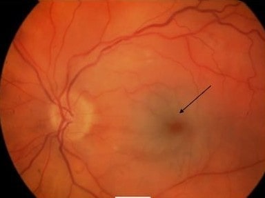
00:00 Welcome back. We are looking at additional diseases that involve the retina and in this particular case we're going to be looking at some important vascular structures, the retinal artery and retinal vein as well as degeneration kind of a primary idiopathic degeneration of the macula. As always, I want to acknowledge the incredible help of Dr. Jose Mata who without whom these images, this slide deck, all the ones you've been seeing and we did it with ophthalmology would not have happened. And so everything you like about this thank Jose; everything you hate about this, let me know. Alright, diseases of the posterior segment, let's talk about retinal vessel occlusion and clearly this is going to be an important thing. So coming out of the optic disc, they're going to be the retinal artery and retinal vein. 00:57 And if they become occluded for a variety of reasons that we'll talk about, ohh hear in a world of hurt, you don't have adequate blood supply into or out of the choroid retina. So risk factors for this are ones that you would probably expect anyway. So patients who are hypertensive will have more atherosclerotic disease, similarly with diabetes more atherosclerotic disease. So these will impact on retinal artery perfusion. Interestingly, atrial fibrillation is also a risk factor and that's probably because embolic disease as we'll see very shortly, embolism into the retinal artery is a major cause of acute retinal artery occlusion. 01:46 So, risk factors were identified, the etiology is embolism, that's why atrial fibrillation with formation of a thrombus in the atrium and then fragmentation and going up into the retinal artery is the most common cause. You can have carotid artery atherosclerosis, you can have fragmentation and embolization of atheroemboli, but you can also have diminished flow in ultimately anterior renal artery. And then there is thrombosis that can happen in the vein or artery for a variety of causes. Usually with these results and as a sudden painless, complete monocular vision loss, that's because there is no blood supply into the eye, into the area of the eye that's going to be transducing light into electrical signals. You can also have very focal visual defects if it's not the main artery, but rather one of the branches. So you can get scotomas, little tiny blind spots around in the visual field. And we'll see how that looks in a minute. So, the retinal pallor here is indicative, on the fundoscopic exam, of diffuse diminution of flow in that portion of the retina and choroid. You can get a cherry red spot that's very typical of a central retinal artery occlusion, it's just kind of an apparent accentuation near the macula. Things are associated with renal vein occlusion are atherosclerotic conditions so diabetes and hypertension are associated with that, but it's not atherosclerosis of the vein, they're just kind of concomitant risk factors. Hypercoagulable state. So retinal vein occlusion can occur when you have sickle cell anemia and you get sludging and then finally occlusion of small vessels due to the abnormal sickle cells. So a hypercoagulable state such as that can cause it. Clearly, inflammation. So if we have a vasculitis associated say with lupus erythematosus, that will cause increased propensity to thrombose in a vein and sometimes pharmacological things such as oral contraception, hyperestrogenic states, cancers, and things such as that may also drive hypercoagulable states that leads to thrombosis of the retinal vein. And then when we occlude the retinal vein, we can't get blood supply out of the retina and choroid and we'll get impressive edema and hemorrhage. So, what are we looking at? This is retinal vein occlusion, this is just the characteristic look to how this occurs, on the left is our normal, on our right we are seeing a variety of things that we should see on a fundoscopic exam associated with this entity. 04:36 Cotton wool spots. Again, cotton wool spots everywhere represent areas of microinfarct. 04:41 We'll also see the so-called blood and thunder appearance. Sounds very dramatic, it's like we should be beating on drums but what is happening is we're getting engorgement of the vasculature and we're getting extravasation of blood so it looks like a little explosion or a big explosion depending on your point of view. There will be papilloedema and engorged retinal veins with associated hemorrhage. So those are kind of the changes we'll see on fundoscopic exam. And again, retinal vein thrombosis leads to a sudden, can be gradual depending on the pace of that thrombosis but largely painless, monocular vision loss.
About the Lecture
The lecture Retinal Vessel Occlusion by Richard Mitchell, MD, PhD is from the course Posterior Segment Eye Diseases.
Included Quiz Questions
What is a risk factor for retinal artery occlusion?
- Diabetes mellitus
- Hypothyroidism
- Cushing's disease
- Multiple myeloma
- Anemia
What is the most common cause of retinal artery occlusion?
- Embolism
- Cerebrovascular aneurysms
- Cranial nerve palsies
- Trauma
- Seizures
What finding is seen on the fundoscopic exam with a retinal ARTERY occlusion?
- Cherry red spot
- Hemorrhage
- Ischemia
- Cotton wool spots
- Copper wiring
What finding is seen on the fundoscopic exam of the retinal VEIN occlusion?
- "Blood and thunder" appearance
- Hard exudate
- Microneovascularization
- Flame-shaped hemorrhages
- Dot-blot hemorrhages
Author of lecture Retinal Vessel Occlusion

Richard Mitchell, MD, PhD
Customer reviews
5,0 of 5 stars
| 5 Stars |
|
5 |
| 4 Stars |
|
0 |
| 3 Stars |
|
0 |
| 2 Stars |
|
0 |
| 1 Star |
|
0 |
