Playlist
Show Playlist
Hide Playlist
Restrictive Cardiomyopathy – Cardiomyopathy

-
Slides 05 Cardiology Alpert.pdf
-
Reference List Cardiology.pdf
-
Download Lecture Overview
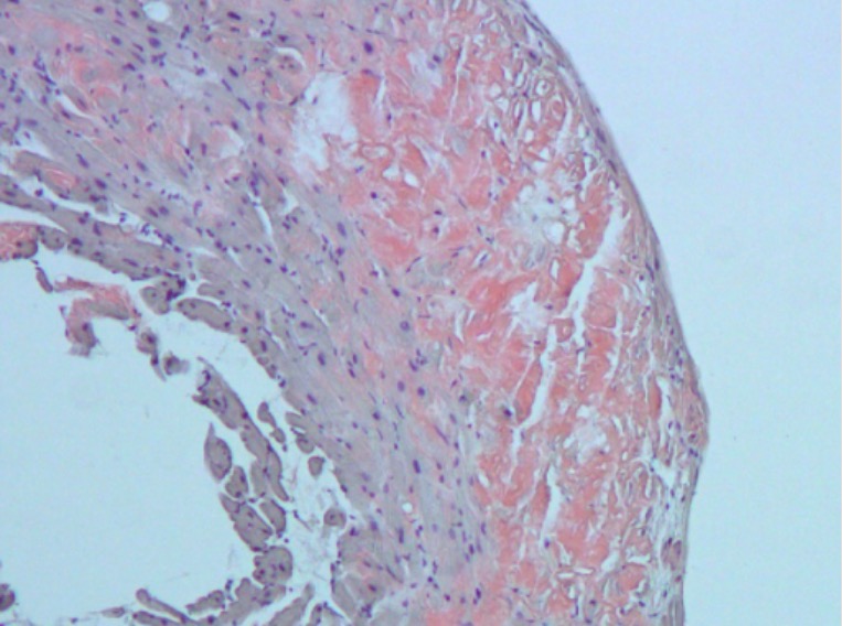
00:00 You don’t, of course, want to remove too much of the septum. 00:04 Well, let’s talk about the much, much less common cardiomyopathies. 00:09 Restrictive cardiomyopathy is almost not seen very much in developed countries. It’s a little more common in developing countries where people have poorer nutritional status. It is a infiltration of the myocardium, usually with a substance that makes the ventricle quite stiff. The commonest one in developed countries is amyloidosis. 00:36 This is an abnormal protein that gets deposited in the heart and makes the heart stiff. And that’s why it’s called restrictive. The heart restricts filling because of this deposition of protein in between the myocardial cells. By the way, amyloidosis can also affect the kidney, so the patients can have kidney failure. It’s a very bad disease, often treated with chemotherapy like a cancer because it’s usually due to a group of immune cells that are producing much too much of an immune substancein the blood and this immune substance gets into the tissues of the heart, the kidney and so forth. It’s called amyloid protein and it causes a marked stiffening of the heart muscle. It has a poor prognosis, again because we first see these patients when they usually have well-developed heart failure; in other words, they’re at a very advanced stage. 01:29 And the treatment - once you try some drugs, the mainstay is diuretics to try and get rid of the excess fluid. Beta blockers, ACE inhibitors don’t work in this form of cardiomyopathy and a number of these patients go on to a heart transplant. These patients can have a lot of arrhythmias as well and can have sudden death. 01:52 Treatment for asymptomatic amyloid related restrictive cardiomyopathy includes treatment for heart failure with diuretics as usual, as well as treatment of the underlying disease. 02:04 For patients with light chain amyloidosis, treatment includes chemotherapy with Mel Fillon plus autologous stem cell transplantation in patients who are not a candidate for stem cell transplantation. 02:17 Autism is a proteasome inhibitor that can be used transpiring. 02:24 Amyloidosis, cardiomyopathy can be treated with taff or meet us, and there may be other drugs coming soon. 02:32 These actually decrease the rate that amyloid is deposited. 02:37 They do not result in resolution and loss of the amyloid that's already there, but they stop further amyloid deposition. 02:45 Patients with this variant of amyloidosis should be evaluated for liver transplantation as well. As well. 02:52 This is in the United States. 02:54 This form of amyloid of transpiring amyloidosis is much more often seen in patients who are African-American. 03:04 Anticoagulation is recommended for any patients with the amyloid cardiomyopathy as well as for patients with atrial fibrillation or ventricular thrombus. 03:15 Now, here you can see the same little diagram we’ve seen before. On the left-hand side is the normal heart and you can see on the right-hand side, there’s a slight thickening of the ventricle. But, what’s really happening here is that the ventricular muscle is being infiltrated with a substance that’s causing it to be restrictive. Sometimes when the pericardium is scarred or injured and gets thickened, it can be hard to distinguish that from a restrictive cardiomyopathy since the physiology is the same - a thickened, stiff pericardium grips the heart and impairs its filling, just as restrictive myopathy does so. And often we need to do MRI and a variety of catheterization tests to decide, “Is this constrictive pericarditis or is this a restrictive myopathy?” Why is that important? Because there’s a surgical operation to remove the pericardium and make the patients much better whereas in restrictive cardiomyopathy, the eventual treatment is heart transplantation. 04:16 This slide just reiterates what I said before. Amyloid is the commonest form in the West of restrictive myopathy. The diagnosis is made usually by echocardiography and the MRI helps us distinguish restrictive myopathy from constrictive pericarditis. This is not a very common thing. In my very busy cardiology hospital, we might see one or two of these, perhaps three patients a year whereas coronary disease is multiple events every day and dilated cardiomyopathy or hypertrophic cardiomyopathy, we see every week. So, you can see a sort of relative experience of the cardiologist. This is not very common. 04:58 And again, treatment - you do the best to keep the patient’s excess fluid on board down with diuretics, but in fact, the prognosis is usually quite poor and these patients may end up, if they are appropriate candidates in other respect, requiring a heart transplant. 05:20 A final type of cardiomyopathy that’s quite rare is called “ARVD” or “arrhythmogenic right ventricular dysplasia” cardiomyopathy. What is this? This is a very unusual form of cardiomyopathy in which the right ventricular muscle is gradually replaced by fat and scar tissue leading eventually to both severe right ventricular failure with symptoms of congestion in the liver and the abdomen and peripheral edema - peripheral swelling and in addition, malignant ventricular arrhythmias that often lead to sudden death. Fortunately, this is a rare cardiomyopathy, but it’s of great interest to scientific investigation because it may give us clues as to why the heart muscle cells are disappearing and dying and perhaps we might be able to reverse it and maybe some of those techniques might be used in other forms of cardiomyopathy. The treatment unfortunately consists mostly of diuretics to control the edema and then implantation of a cardioverter–defibrillator. So, when the patient develops a malignant ventricular arrhythmia, they will be shocked out of it and will not have a sudden death. 06:34 A number of these patients go on to, of course, heart transplant. It’s genetically transmitted in families, and as I said, even though it’s rare, there’s a great deal of scientific interest in this particular form of cardiomyopathy. What happens here in this rare form of cardiomyopathy is something that’s of great interest to scientists who are studying the forms of cardiomyopathy. 06:58 These individuals are usually born normal. They develop normally and then as they age, progressively the right ventricular heart muscle is replaced by fat and scar tissue. 07:10 It’s a genetic cardiomyopathy. The underlying mechanism is not totally understood, but what happens is these patients go on to develop severe right ventricular failure. Remember, that’s with elevated neck veins, fluid in the abdomen, fluid in the legs. The left ventricle is often normal although occasionally affected, but it’s mostly the right ventricle that’s affected. 07:33 And what happens is these patients develop malignant ventricular arrhythmiasand sudden death and they often need treatment not only for their heart failure, but also they need an implantable defibrillator. In case they develop ventricular fibrillation cardiac arrest, it will shock them out of it. It’s very uncommon, but in fact, is the source of a lot of scientific investigation because if we could understand some of the mechanisms - why the heart cells disappear and fat takes their place, it might help us to understand what’s going on in some other forms of cardiomyopathy and we might even be able to come up with some innovative therapies. It’s a dominant autosomal inherited disease. 08:19 The electrocardiogram has a specific finding that helps you to identify it, but usually, the best way is with MRI where we can actually see the fat replacing the right ventricular muscle in arrhythmogenic cardiomyopathy. Let me just give you some final hints about
About the Lecture
The lecture Restrictive Cardiomyopathy – Cardiomyopathy by Joseph Alpert, MD is from the course Cardiac Diseases.
Included Quiz Questions
What is a cause of restrictive cardiomyopathy?
- Amyloidosis
- Viral infection
- Dietary protein deficiency
- Bacterial infection
- Trauma
Arrhythmogenic right ventricular dysplasia (ARVD) is the 2nd most common cause of sudden cardiac death in young people after hypertrophic cardiomyopathy. What is the best diagnostic tool for ARVD?
- MRI
- CT
- Ultrasound
- PET/CT
- X-ray
Author of lecture Restrictive Cardiomyopathy – Cardiomyopathy

Joseph Alpert, MD
Customer reviews
5,0 of 5 stars
| 5 Stars |
|
5 |
| 4 Stars |
|
0 |
| 3 Stars |
|
0 |
| 2 Stars |
|
0 |
| 1 Star |
|
0 |
