Playlist
Show Playlist
Hide Playlist
Lymphadenopathy: Reed-Sternberg Cells – White Blood Cell Pathology


-
Slides Lymph Nodes.pdf
-
Download Lecture Overview
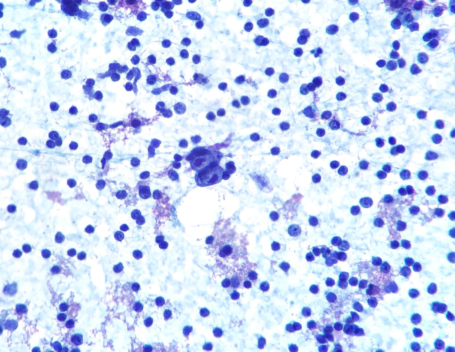
00:00 Upon morphology of Hodgkin lymphoma, what you want to do is with this illustration or these illustrations, you compare these with the previous discussion that we had on the table, where you have the different types of morphology of what? The topic for this entire section is morphology of Reed-Sternberg cell. 00:21 You must find the Reed-Sternberg cell so that you can do what? Diagnose Hodgkin. 00:26 If you do not find an owl eye type of appearance upon histology and your patient has B symptoms. 00:33 What do B symptoms mean to you? Night sweats, weight loss and you have fever. 00:38 Sounds an awful lot like TB, but B symptoms could be found both on Hodgkin and non-Hodgkin. 00:44 Reed-Sternberg cell, only Hodgkin. 00:47 The classic type, take a look at this. 00:50 And it looks like owl eyes, doesn’t it? You have the nucleoli which is looking right back at you. 00:55 Then you have the lacunar type. 00:57 I want you to pay closer attention to the lacunar and the one that you’re paying attention to is the one in the upper left corner. 01:04 And you find a lot more space or vacuolization, that’s a lacunar type. 01:10 Remember that classic and lacunar could be found with the sclerosing type of Hodgkin, being the most common. 01:18 And I want you to move down to the mononuclear. 01:20 With the mononuclear, what looks like a mononucleus and, in addition, the mononuclear variant will be found with mixed more so cellularity type of Hodgkin, M and M. 01:32 And then bottom right, you find your popcorn cells or L&H (lymphocytic and histiocytic) and this type of Reed-Sternberg cell will be found with nodular lymphocyte predominant type. 01:47 Four different variants of Reed-Sternberg cell. 01:50 If I were you, I’d be very comfortable with being able to identify each one of these types of Reed-Sternberg cell based on the type of Hodgkin that your patient is presenting with. 02:03 Now the Reed-Sternberg and morphology, the description. 02:07 The classic Reed-Sternberg cells are large. 02:11 We'll walk through the description. 02:13 With abundant pale cytoplasm and two or more oval lobulated nuclei containing prominent “owl eye” eosinophilic nucleoli. 02:25 I will pause here for one second and make sure that you repeat this a few times to yourself. 02:31 So that when you read this in a stem of a question, that you will know that you’re referring to the Reed-Sternberg cell. 02:37 They might not, at all times, come out and say Reed-Sternberg, but they will go ahead and tell you that there is quite a bit of cytoplasm and you have these oval lobulated nuclei giving you the owl eye with eosinophilic nucleoli. 02:54 In some Reed-Sternberg cell variants, the cytoplasm shrinks and due to this, it then appears as though that you have quite a bit of emptiness or vacuolization around the nucleus. 03:07 Don’t worry about the mechanism of the shrinkage, that’s not important. 03:13 Be able to identify a lacunar cell. 03:16 So there is going to be increased emptiness or vacuolization around the nucleus. 03:21 And I’ve shown you picture in the previous discussion. 03:24 Make sure you know the description of lacunar and your classic. 03:29 And then you have the L&H. 03:31 The L&H, if you go back and take a look at the picture, is fluffy. 03:35 It looks lobulated and looks like popcorn, hence L&H. 03:42 Here, this type of variant is found with nodular lymphocyte predominant. 03:47 Good. 03:48 Not lymphocyte-rich. 03:49 Keep those separate. 03:55 In this section, we’ll have to walk you through Ann Arbor classification in great detail. 04:01 Every single stage, you must know because of Hodgkin and because of prognosis. 04:08 Let’s begin with stage I. 04:10 Stage I is one lymph node affected. 04:14 The specific classification is called Ann Arbor, only for Hodgkin here, not non-Hodgkin. 04:20 One lymph node affected and if it’s something like your sclerosing type, may be the mediastinum or the patient said, “Doc I have a lump in my neck.” Does it hurt? "No." Oh, boy. 04:32 Your concern upon biopsy, you find a Reed-Sternberg cell. 04:38 A nuclei that are lobulated, looking back at you, “owl eyes”. 04:42 And in addition, you find a classic type or a lacunar. 04:47 You might be thinking about nodular sclerosing especially if it’s a female. 04:52 Stage II, you have two or more lymph nodes that are affected, but only one side of the diaphragm. 04:58 Locate the diaphragm to yourself please And on one side of diaphragm you have 2 lymph nodes that are affected. 05:03 Maybe the cervical or maybe the mediastinal. 05:06 Obviously, they are nontender. 05:09 Now, we go from stage II to stage III and this is a pretty extensive change. 05:15 What happens now is the fact that you have two or more lymph nodes on either side of the diaphragm. 05:22 So if by chance you find involvement of your, let’s say, mediastinal lymph node and then you find involvement of the spleen. 05:30 Spleen. 05:33 From henceforth, you must think of the spleen pathologically as being a lymph node. 05:36 Please do that. 05:38 So therefore if you have one lymph node that is affected by the mediastinum and you have another lymph node that has affected “the spleen” that will be the other side of the diaphragm, that is 2 or more, so you satisfied that criteria, in addition, you’re on either side of diaphragm, both sides of diaphragm. 05:55 This is not stage I, definitely not stage II. 05:58 In fact this is stage III. 05:59 You’re on both sides of the diaphragm. 06:02 Once you’re at stage IV, unfortunately, extralymphatic spread, metastasis. 06:06 But even with this though, prognosis pretty decent. 06:11 These are stages of Hodgkin lymphoma. 06:15 Spend a little bit of time especially with stage II and stage III so that you understand as to when you've moved from stage II to stage III depending as to what side of diaphragm you’re on, or on one side or both sides. 06:30 Clinical presentation of Hodgkin, the B symptoms include once again, what are they? Fever, weight loss and night sweats. 06:38 We have gone over that over and over again. 06:39 At this point, I expect you to know what that is. 06:42 Do not confuse this with TB, tuberculosis. 06:46 Prognosis depends on stage, but even with the later stages, what is the name of staging mechanism of Hodgkin? Good. 06:56 It’s called Ann Arbor. 06:57 It’s still generally good. 07:00 Or even stage IV, take a look at this. 07:02 Very rarely can you get to stage IV in a cancer and have such decent, decent type of survival rate, 60%-70% 5-year survival. 07:14 And it’s important that you know the CD markers. 07:18 Go in 15s. 07:21 Pay attention to 15 and 30 and 45; 15 positive, 30 positive, 45 negative; 15, 30, 45. 07:30 Memorize the 15 and 30 will be positive and 45 will be negative. 07:34 Those three for sure, you need to make sure that you know. 07:37 All of your Hodgkin lymphoma will be of that pattern of cluster differentiation except for the popcorn. 07:46 All right. The popcorn was the one that we talked about L&H. 07:49 And that is the one that is called nodular lymphocyte predominant. 07:53 And that will be one in which 15, 30 negative and 45 positive. 07:57 But your focus should be on 15, 30 positive for sure.
About the Lecture
The lecture Lymphadenopathy: Reed-Sternberg Cells – White Blood Cell Pathology by Carlo Raj, MD is from the course Lymphadenopathy – White Blood Cell Pathology (WBC).
Included Quiz Questions
What is the characteristic appearance of Reed-Sternberg cells?
- Abundant, pale cytoplasm, and two or more oval lobulated nuclei containing prominent owl's eyes eosinophilic nucleoli.
- Abundant, pale cytoplasm, and two or more circular lobulated nuclei containing prominent owl's eyes basophilic nucleoli.
- Scarce, pale cytoplasm with one or more semicircular portions removed from the cell margin and a prominent owl's eyes basophilic nucleoli.
- Scarce, pale cytoplasm, and one oval lobulated nucleus containing prominent owl's eyes basophilic nucleoli.
- Abundant, pale cytoplasm with one or more semicircular portions removed from the cell margin and a prominent owl's eyes eosinophilic nucleoli.
Which of the following describe stage III of Ann Arbor staging for lymphoma?
- Two or more lymph nodes on both sides of the diaphragm
- One lymph node on the same side of the diaphragm
- Two lymph nodes on the same side of the diaphragm
- One lymph node each, on both sides of the diaphragm
- Two lymph nodes on the same side of the diaphragm along with extranodal involvement
Which of the given CD markers are positive in the nodular lymphocyte-predominant form of Hodgkin lymphoma?
- CD20, CD45
- CD11, CD91
- CD3, CD117
- CD15, CD30
- CD34, CD41
Author of lecture Lymphadenopathy: Reed-Sternberg Cells – White Blood Cell Pathology

Carlo Raj, MD
Customer reviews
5,0 of 5 stars
| 5 Stars |
|
5 |
| 4 Stars |
|
0 |
| 3 Stars |
|
0 |
| 2 Stars |
|
0 |
| 1 Star |
|
0 |
