Playlist
Show Playlist
Hide Playlist
Rectus Sheath

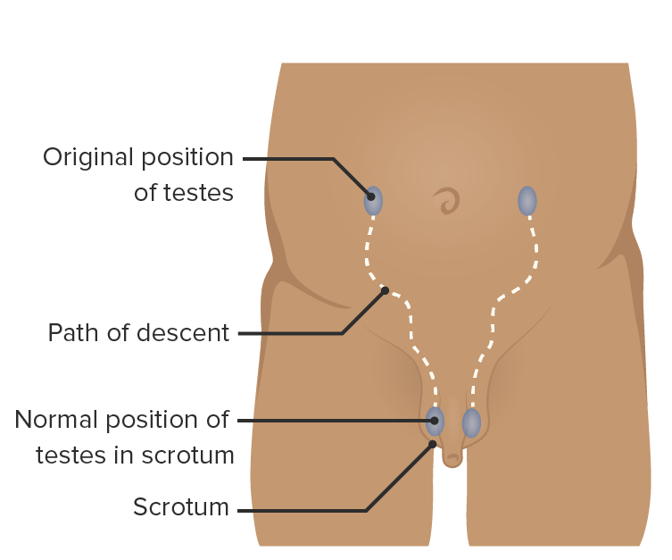
00:00 You imagine if I make a paramedian incision here, what are the things I’ll go through? Skin, subcutaneous fat, Camper’s, Scarpa’s. 00:12 Before that? Sheath, rectus sheath. 00:16 What is the rectus sheath made of, here? The anterior leaf of the internal oblique. 00:28 So, you have linea alba. 00:31 That's your rectus abdominis. This is umbilicus. 00:38 You have the external oblique coming here, internal oblique, and transverse abdominis. 00:46 The external oblique comes, aponeurosis goes in front. 00:52 The internal oblique splits into two. 00:55 That's the anterior leaf. This is the posterior leaf. 01:00 This is the transverse abdominis. 01:03 If you are making an incision through there, you are going through the external oblique, the anterior leaf of the internal oblique, rectus abdominis, posterior leaf of the internal oblique, aponeurosis of the transverse abdominis, transversalis fascia. 01:22 We have transversalis fascia everywhere, then preperitoneal fat, and peritoneum. 01:29 What's the difference between that? And if I make an incision lower down here, will it be the same? What's the difference? There is no posterior rectus sheath. 01:42 Below the arcuate line which is midpoint between the pubic symphysis and the umbilicus there, at this point, it's called the arcuate line. 01:56 Below that, there is no posterior rectus sheath. 02:00 You only have the anterior rectus sheath. 02:02 All these three layers essentially go in front. 02:10 What's the clinical significance of that? What's the clinical significance of no posterior rectus sheath there? The clinical significance is that is the weakest area in the abdomen. 02:22 That's where you get a direct herniation because that is weak. 02:26 There's no posterior rectus sheath. 02:28 Your direct hernia is a protrusion in the transversalis fascia just pushing out because there is no sheath to protect it below the arcuate line. 02:39 That brings us to, as we see there, midline, rectus muscle on either side, umbilicus, pubic symphysis. 03:04 If I take this line, anterior superior iliac spine, pubic symphysis, pubic tubercle, that's your inguinal ligament. 03:17 In the mid-inguinal point is a midpoint between the pubic symphysis and the anterior superior iliac spine, you have the femoral artery. 03:33 Sorry, I should have drawn that in red, femoral artery. 03:42 You have that deep inguinal ring between the pubic tubercle and the ASIS which is called the midpoint of the inguinal ligament which is here. 04:03 Go on. Is there anything you want to ask? It’s okay. 04:06 That’s okay. 04:08 Go on. 04:09 I kind of missed what the difference was below the arcuate line. 04:12 I'm sorry. 04:13 No, that's fine, below the arcuate line. 04:18 Imagine this is your rectus abdominis, above the arcuate line, you have the aponeurosis going in the front and then behind. 04:30 This is essentially rectus sheath. 04:32 Below the arcuate line, all the aponeurosis are going in the front. 04:35 That means there is no posterior rectus sheath. 04:39 That means the posterior rectus, that area becomes weaker because there's no posterior sheath. 04:48 From here, you have the inferior epigastric artery running to the lateral border of the rectus muscle which essentially forms your Hesselbach's triangle. 05:08 Hesselbach's triangle is bounded medially by the lateral edge of rectus abdominis, laterally by the inferior epigastric artery, and inferiorly by the inguinal ligament. 05:21 This is the weak spot for herniation because there is no posterior rectus sheath. 05:27 Staying on here above the inguinal canal is there. 05:35 Approximately 1.25 to 1.50 centimeters above the inguinal ligament is where you have the deep inguinal ring. 05:45 If you have an inguinal hernia, it enters the deep inguinal ring this way and comes into the scrotum. 05:51 Direct hernia will never ever come into the scrotum. 05:55 Direct hernia will have to stop in the inguinal region. 05:58 It can't come into the scrotum anatomically. Okay? Right.
About the Lecture
The lecture Rectus Sheath by Stuart Enoch, PhD is from the course Upper Part of the Body Anatomy.
Author of lecture Rectus Sheath

Stuart Enoch, PhD
Customer reviews
5,0 of 5 stars
| 5 Stars |
|
5 |
| 4 Stars |
|
0 |
| 3 Stars |
|
0 |
| 2 Stars |
|
0 |
| 1 Star |
|
0 |
