Playlist
Show Playlist
Hide Playlist
Pupillary Control – Physiology Review

-
Slides Optic Pathology Pupillary Control and Disorders.pdf
-
Reference List Pathology.pdf
-
Download Lecture Overview
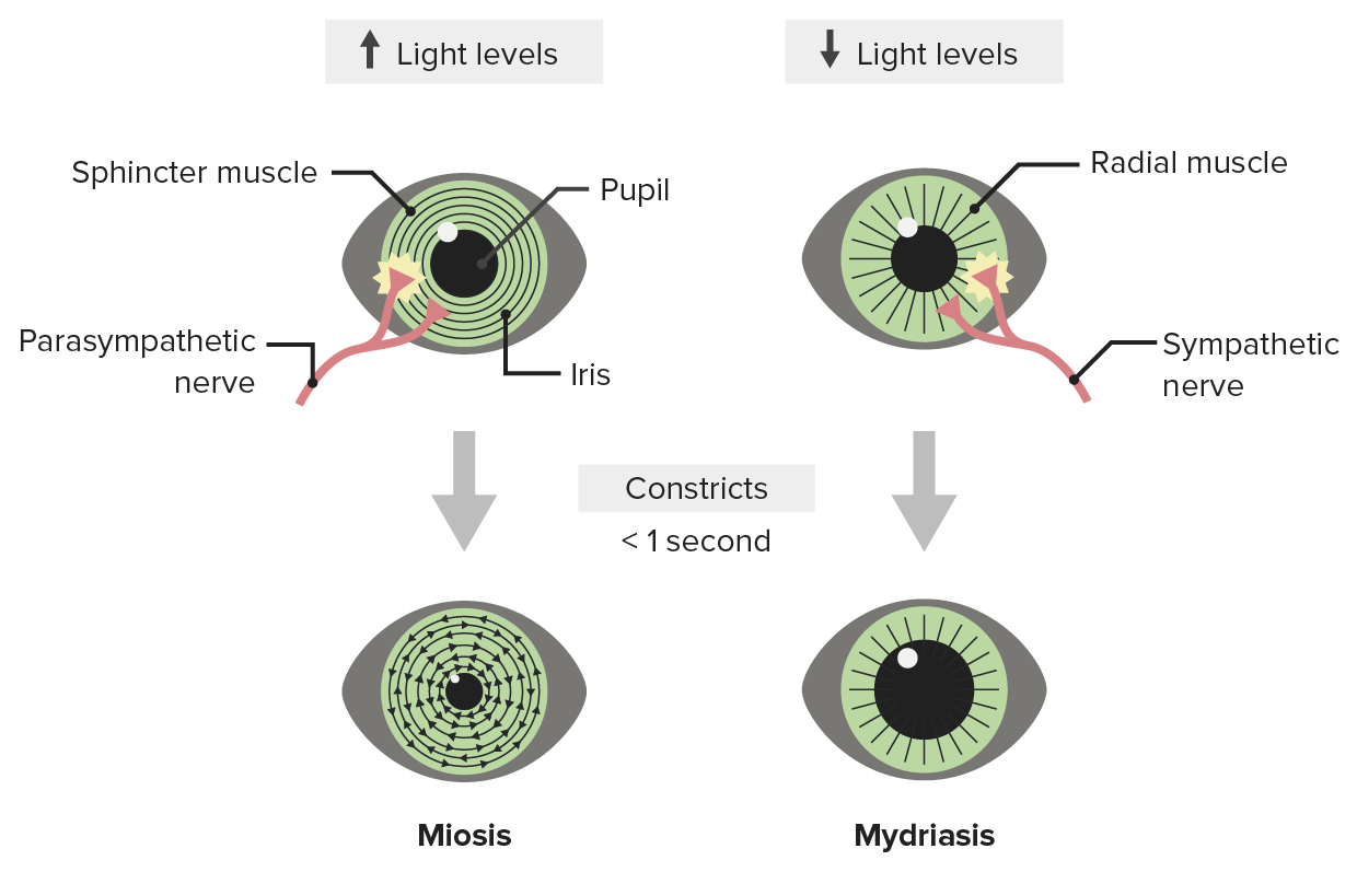
00:02 Alright, as we continue our crusade through various pathologic disorders involving the anterior chamber and uvea, we're going to look at how we control the pupil's, pupillary control, whether they constrict or dilate. 00:15 And some of the disorders, which are kind of interesting. 00:19 All of this is interesting, of course, but this is particularly really cool. 00:22 Alright. 00:24 So this is where we're looking at, we're going to be paying attention to how the iris moves. 00:28 Remember, the iris is not only heavily pigmented to limit light to just going through the pupil but it's also got a fairly high investment of smooth muscle and the smooth muscle is in two concentric rings, there are some that is radially distributed that allows the eye to dilate. 00:48 And then there are some that is concentrically oriented, allows it to constrict much like a camera diaphragm. 00:54 Okay. 00:55 So a normal pupil is that. 00:58 A constricted pupil is called miosis. 01:01 And that is that inner circular layer that is undergoing a contraction of the sphincter pupillary muscle. 01:09 The Innervation is parasympathetic. 01:12 So this is driven through the acetylcholine receptors, particularly the muscarinic receptors. 01:18 Okay, so that's a constricted pupil. 01:20 On the other end of the equation is the dilated pupil, or mydriasis. 01:25 And the mydriasis is a contraction of the dilator pupillary muscle, which is radially oriented. 01:30 So you can see the little arrows all the way around which will pull the pupil open by causing contraction of the smooth muscle cells there in the iris, that innervation is sympathetic. 01:42 So this has implications. 01:44 When you are excited, your pupils are dilated because of epinephrine. 01:49 And we can also cause dilation of pupil through a variety of drugs, including many that are illicit. 01:56 But tropicamide is a compound that we put into the eye that will actually be a muscarinic receptor antagonist and antagonizes the miosis and the eyes dilate. 02:09 We do that so that we can get nice wide open pupils. 02:13 And we can see into the fundus. 02:15 And so when you go for your eye exam, they will typically put some of that topical tropicamide into your eye. 02:21 And we'll get a nice dilated pupil, that's also going to make you a little photophobic because you can't contract it for about a half hour afterwards. 02:30 One other interesting historical point, it used to be thought back in the Renaissance and in some of the areas around that period of time that women with big dilated pupils were beautiful, it was a sign of beauty. 02:44 And so they learned or was discovered is that you could put a little belladonna, which is a muscarinic receptor antagonist in your eye and it would dilate up. 02:52 Of course, you couldn't see a damn thing because you're very photophobic, but you look beautiful. 02:58 Alright, so now you know much more than you wanted to know about miosis and mydriasis. 03:03 How is this working? How do we get? So there are different things that we also need to consider here. 03:09 In terms of the neuronal control of this because when one eye constricts to a light, usually the other one does too. 03:18 So there's a pupillary light reflex. 03:20 So we have a right eye light stimulus, indicated in green. 03:26 And that's going to travel down the optic nerve going all the way back into the superior colliculus, that's the segment of brain that's described there. 03:33 So this is kind of high midbrain. 03:36 And in the superior colliculus, that information that light is coming in through the right eye come in through the optic nerve is transmitted to the pretectal nucleus. 03:47 And then... 03:49 where we get now combined information to both eyes it goes out to the Edinger-Westphal nucleus, which then has tracks that go out along the oculomotor nerve. 03:59 So cranial nerve number three, that will impact on or impinge on the ciliary ganglia and cause constriction. 04:08 And not only that, but it's constriction in both eyes, even though one is being stimulated. 04:12 So we see miosis in both eyes, that's the normal pupillary light reflex. 04:18 Beautiful. 04:19 Rewind that again and run through it, but it's really cool. 04:22 Now we're going to mess that up in a couple different ways.
About the Lecture
The lecture Pupillary Control – Physiology Review by Richard Mitchell, MD, PhD is from the course Diseases of the Anterior Chamber and Uvea.
Included Quiz Questions
What is the densely pigmented region of the eye called?
- Iris
- Cornea
- Conjunctiva
- Pupil
- Macula
What are the types of smooth muscles present in the iris?
- Radial and concentric
- Straight and diverging
- Coarse and fibrillar
- Fine and tortuous
- Elastic and fibrous
What does the parasympathetic stimulation of the sphincter pupillae muscle cause?
- Miosis
- Mydriasis
- Photophobia
- Diplopia
- Hyperopia
What is dilation of the pupil called?
- Mydriasis
- Miosis
- Diplopia
- Myopia
- Emmetropia
Tropicamide is a...
- ...muscarinic antagonist.
- ...muscarinic agonist.
- ...adrenergic antagonist.
- ...beta blocker.
- ...nicotinic agonist.
Author of lecture Pupillary Control – Physiology Review

Richard Mitchell, MD, PhD
Customer reviews
5,0 of 5 stars
| 5 Stars |
|
5 |
| 4 Stars |
|
0 |
| 3 Stars |
|
0 |
| 2 Stars |
|
0 |
| 1 Star |
|
0 |
