Playlist
Show Playlist
Hide Playlist
Pulmonary Embolism: Pulmonary Contrast Angiogram

-
Slides 11 VascularMedicine advanced.pdf
-
Reference List Vascular Medicine.pdf
-
Download Lecture Overview
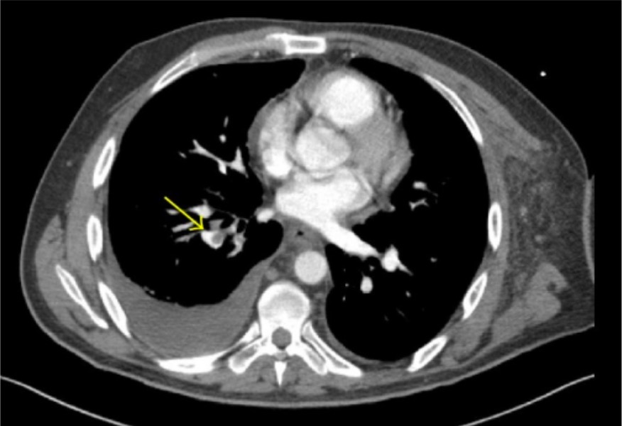
00:00 These days, the diagnosis of pulmonary embolism is almost always made by a CT angiogram in which we actually see contrast arriving in the artery and showing that there’s a defect where the clot is. 00:19 This is actually a contrast pulmonary angiogram where a catheter is inserted and squirts the dye in. But in fact these days the CT scans are so accurate that we can just administer the dye intravenously, do the CT scan, see it when it arrives in the lung bed and pick up the pulmonary embolism. This test has now essentially replaced the lung scan except in patients who are allergic to the angiographic dye. Those patients may still get a lung scan and we accept in the clinical setting that the lung scan is not as accurate as the CT angiogram. 00:54 The CT angiogram is high accuracy in the case where we put a catheter in. It’s an invasive test so there’s a chance for complications of the invasive test. But with the CT angiogram, it’s mostly a non-invasive test, just with some intravenous injection of the dye. 01:12 And here you see a very nice picture showing a beautiful clot in the inferior interlobar branch of the left pulmonary artery. 01:21 You can also do an angiography by MRI. Here you see an MRI angiogram and, again, the green arrow points out a clot in this MRI angiogram. By the way, you can also see the aorta and the brachiocephalic artery and the left subclavian and the left carotid artery as well. 01:42 You can see how beautifully visualised the arteries are with this MRI. And the CT angiogram gives you a similar picture. Very, very excellent picture, both non-invasive.
About the Lecture
The lecture Pulmonary Embolism: Pulmonary Contrast Angiogram by Joseph Alpert, MD is from the course Venous Diseases.
Included Quiz Questions
Which one of the following regarding CT angiogram is NOT true?
- It does not need an angiographic dye.
- It has essentially replaced the lung scan.
- It is considered to be noninvasive when performed with IV contrast.
- It has high accuracy.
- It can be successfully used to diagnose a clot in the pulmonary artery.
For patients allergic to contrast material the noninvasive procedure done to see pulmonary embolism may be?
- MR angiography
- Spirometery.
- Chest X-ray.
- Blood CP.
- CT scan.
Author of lecture Pulmonary Embolism: Pulmonary Contrast Angiogram

Joseph Alpert, MD
Customer reviews
5,0 of 5 stars
| 5 Stars |
|
5 |
| 4 Stars |
|
0 |
| 3 Stars |
|
0 |
| 2 Stars |
|
0 |
| 1 Star |
|
0 |
