Playlist
Show Playlist
Hide Playlist
Pulmonary Embolism: Clinical Findings


-
Slides PulmonaryEmbolism RespiratoryPathology.pdf
-
Reference List Pathology.pdf
-
Download Lecture Overview
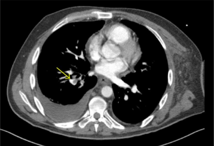
00:01 Alright, with pulmonary thromboemboli, let's take a look at the symptoms of your patient. 00:04 The clinical findings include the following - well in the very beginning of this lecture I was trying to demonstrate the increased breathing, wasn’t I? And I was doing rapid shallow breathing. 00:15 Now what does that mean to you? Then you bring in once again your physiology remember that all important alveolar ventilation we've talked about, and what does that mean to you? As your tidal volume multiply that by the respiratory rate but before you get to alveolar ventilation, what must you then subtract? Oh, remember that oxygen that’s playing hide and seek? What's hiding, what’s it called? Dead space - so therefore you have your tidal volume and from this you subtract your dead space approximately 150, and then you multiply this by the frequency or your respiratory rate. 00:46 We'll talked about this over and over again and all I'm doing is just properly incorporating it. 00:50 So imagine now that you have rapid shallow breathing so now I have my tidal volume being approximately or maybe it’s all the way down to 300. 00:59 Three hundred from this you subtract your 150 multiply that by the respiratory rate, you don’t get a lot of alveolar ventilation so therefore, maybe, you might have issues with hypoxia, yeah, you will. 01:11 And maybe perhaps even maybe, maybe - have issues with carbon dioxide not being effectively blown off, just keep that in mind. 01:18 Rapid, shallow, not necessarily a good thing. 01:21 What else is going to happen? Well, you have your tachycardia, tachypnea and chest pain and, well the differentials are still broad in that area you still have your lung, you have your heart and you have your, well, COPD so you have your pneumonia, heart failure and such. 01:36 If you are suspecting a patient with any type of heart failure then as soon as they walk in through the door what are you gonna do with that patient who is at this point having issues with breathing? How about an ECG just to make sure that we rule out anything from the heart causing damage to the lung? And if the ECG is perfectly normal, you don’t find any ST elevations, no Q waves, proper sinus rhythms, so on and so forth we can then well maybe not think of heart being an issue but keep in mind though if you do have lung issues, then you would expect to see issues where though? More so on the right side, so you might be able to find on your ECG more of a right ventricular hypertrophy, you call this right-sided strain, or right ventricular strain and you might find evidence of what's known as right atrial enlargement. 02:22 Are we clear? If it’s lungs that’s being affected therefore right side of the heart. 02:26 If it's pneumonia, then you're thinking about looking at the chest x-ray and you’ll find increase opacity perhaps, if you're thinking about lobar, lobar - but if you find really no or it’s unremarkable, and all that you find is just issues with the blood vessels and such, less likely to be pneumonia. 02:44 And then COPD exacerbations, well, that’s relatively straight forward because that would then mean a whole different set of test such as your pulmonary function tests you would find your FEV 1 to FVC ratio to be depressed and you're on your merry way. 02:57 Let’s continue. 02:59 Patients with DVT often have signs and symptoms of LE stands for? Lower extremity DVT. What does that even mean? That means that upon dorsiflexion, can you do that for me right now? You're lifting your foot? Good. 03:12 And upon your dorsiflexion you end up having pain perhaps in the back of the leg by the calf and that may then give you a clue. 03:20 Remember, you don’t always have to find that, is that clear? Now, if you see it, and you read it, and you hear it, highly differential would be DVT. 03:28 Now, What about the clinician? Well, you have just - well, you're responsible for doing rounds, right? And during your rounds here we are and then we're seeing a patient who's been lying in this bed for quite some time, hospitalized patients, cancer. 03:45 These patients are extremely hypercoagulable especially if they are nonambulatory. 03:52 Therefore what you might wanna do that you can just prevent a DVT from developing? How about a little walk around the block so you're not gonna ask your patient to run 10 miles, they just had surgery. That’s not what I mean. 04:07 You just take them up and they're stuck with their IV, don’t pull them too hard, that will hurt too, so you just slowly walk him around the hall whatever and trust me makes a world a difference. 04:18 Or maybe what else might you wanna do? How about some hosiery, how about some compression socks on your legs so that you can prevent that DVT from developing? Big time important time, big time - because you don’t want a patient that dies on your watch.
About the Lecture
The lecture Pulmonary Embolism: Clinical Findings by Carlo Raj, MD is from the course Disorders of the Pulmonary Circulation and the Respiratory Regulation: Basic Principles with Carlo Raj.
Included Quiz Questions
Which of the following findings would you expect on the echocardiogram of a patient with a large pulmonary embolism?
- Right ventricular enlargement
- Elevated left ventricular pressure
- Decreased left atrial pressure
- Decreased right atrial pressure
- Left ventricular enlargement
A patient presents with shortness of breath, tachycardia, tachypnea, and pleuritic chest pain. Other than pulmonary embolism, what differential diagnosis should be urgently considered?
- Myocardial infarction
- Myocarditis
- COPD
- Pneumonia
- Endocarditis
Author of lecture Pulmonary Embolism: Clinical Findings

Carlo Raj, MD
Customer reviews
5,0 of 5 stars
| 5 Stars |
|
5 |
| 4 Stars |
|
0 |
| 3 Stars |
|
0 |
| 2 Stars |
|
0 |
| 1 Star |
|
0 |
