Playlist
Show Playlist
Hide Playlist
Proteus Mirabilis

-
01-18 Proteus Mirabilis.pdf
-
Download Lecture Overview
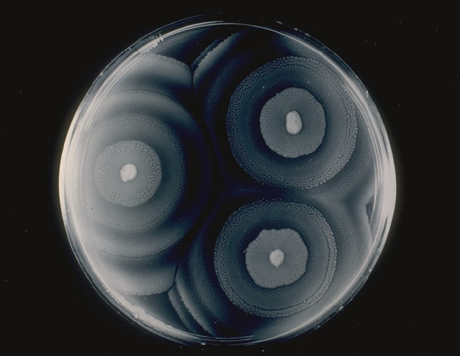
00:01 Proteus Mirabilis, a bacteria. 00:04 Proteus are gram-negative rods which are none lactose fermenting and have a strong urease activity. 00:12 They also have a swarming growth on agar, which is one of their unique features. 00:19 The proteus typically colonize the GI-tract. 00:24 Where they live quite happily and normally don't cause any disease whatsoever until or if they're transmitted to the urinary collecting system, the urinary tract via spread of fecal flora. 00:37 Now, this can occure accidentally and in fact most commonly is an accidental transmission. 00:42 Sometimes however there can be introduction via introduced specimens such as retained toilet paper which can also cause an ascending infection. 00:53 How does this organism cause its disease? Well, it basically creates an alkaline urine and it does so through its very strong urease activity. 01:06 -ace, an enzyme which cleaves urea into ammonia and carbon dioxide. 01:13 That combination of by-products increases the pH of the urine allowing the organism to survive. 01:20 However, it unfortunately also increases the collection of renal stones because many of the stone materials will silt out, or become solid at that particular pH. 01:33 And a higher increased urine pH is quite toxic to the uroepithelium. 01:40 So, proteus mirabilis which has a protease is a common cause of urinary tract infections and along with those associated staghorn calculus formation. 01:55 Also, because of the urease which it contains and it creates ammonia, you can often tell that a patient has a urinary tract infection with proteus because the urine smells strongly of ammonnia. 02:08 This image shows two side to side comparisons of cystourethrogram of a patient with a staghorn calculus. 02:17 If one looks at the initial image on the left of side of the screen, one can see on the right side of the patient or the right side of the image which is the left side of the patient, a large white structure which looks sort of a globoid. 02:34 That would be your staghorn calculus. 02:37 The image on the right now shows further escalation of the dye from the cystourethrogram outlining the kidney on the patient's right or the image's left and showing significant dilation of the intranefric or the intra-kidney structures. 02:55 This typically would come from obstructive hydronephrosis created by calculi further on down. 03:02 So, this patient is an unfortunate, very clear example of formation of a staghorn calculus. 03:08 Why staghorn? Well, as a stag with a rack of antlers you can see multiple elements projecting into the renal parenchyma which are all calcified or solidified formations of that renal stone. 03:23 Looking just like a rack of antlers. 03:25 On this slide, the image on the left is another example of an intravenous pyelogram ivp showing a staghorn calculus prior to surgery. 03:33 The stones are composed primarily of struvite and carbonic appetite and usually contain many bacterial colonies. 03:40 The image on the right shows the specimen removed from the patient in surgery. 03:44 And here you can you can appreciate both the size 123 grams and the staghorn shape. 03:51 Treatment for this organism. 03:53 Most often the organisms are ampicillin susceptible or penicillin susceptible but so too one can use Trimethoprim/Sulfamethoxazole, sulfonamide, or cephalosporins. 04:04 So, what to do about this particular organism? Well, I'm sorry to say if you find it smelling like ammonia and creating calculi then you've got your diagnosis even before waiting for the beautiful culture of a swarming bacteria.
About the Lecture
The lecture Proteus Mirabilis by Sean Elliott, MD is from the course Bacteria.
Included Quiz Questions
What is the pattern of growth of Proteus mirabilis on a culture plate?
- Swarming growth on agar
- Filamentous growth on agar
- Spreading growth on agar
- Mucoid growth on agar
- Powdery growth on agar
Which of the following is a typical finding in infections caused by Proteus mirabilis?
- Ammonia smell in urine
- Fruity smell in urine
- Proteins in urine
- Glucose in urine
- Foul smell in urine
Author of lecture Proteus Mirabilis

Sean Elliott, MD
Customer reviews
5,0 of 5 stars
| 5 Stars |
|
5 |
| 4 Stars |
|
0 |
| 3 Stars |
|
0 |
| 2 Stars |
|
0 |
| 1 Star |
|
0 |
