Playlist
Show Playlist
Hide Playlist
Plantar Aspect – Anatomy of the Foot

-
Slides 07 LowerLimbAnatomy Pickering.pdf
-
Download Lecture Overview
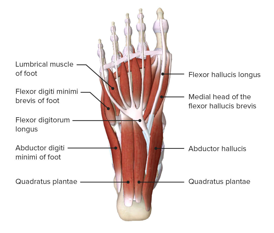
00:01 Now let’s turn to the plantar aspects of the foot and the plantar aponeurosis and its various compartments. So what we see is if you take away the skin of the foot, you have a very tough membrane, the tough connective tissue layer, and this is known as the plantar aponeurosis. We can see it here. It’s thickened within this central compartment of the foot, just like the palmar aponeurosis in the hand. So the deep fascia of the foot, it passes down from the deep fascia of the leg. And dorsally, we can’t see it here on the screen, but it’s very thin and it’s continuous with the extensor retinaculum. But on the sole of the foot, the deep fascia is continuous with the plantar fascia. And as I said, this is thickened centrally as the toughened plantar aponeurosis. This is really important. 00:55 This tough plantar aponeurosis, we can see it here. This tough plantar aponeurosis, this triangular-shaped thickening of aponeurosis, this plantar aponeurosis protects the sole of the foot from injury. 01:13 As we’ll see later, it supports longitudinal arches of the foot. So it helps to maintain those arches and it helps to hold the bones of the foot together. So it’s a really important membrane, this plantar aponeurosis. So extending from the calcaneus all the way towards the digits, the plantar aponeurosis actually divides into five bands. And these contain the flexor tendons of the digits. At the level of the heads of the metatarsals, the plantar aponeurosis, as we can see here, is actually reinforced, and it forms its ligament, which is known as the superficial transverse metatarsal ligament. And this helps to hold the metatarsals together. 02:01 Passing superior are intermuscular septa and these divides the sole of the foot into compartments. 02:08 So the plantar aponeurosis is most inferiorly, and then passing superiorly up through the foot of a whole series of these intermuscular septa, and they divide the foot into compartments, a lateral, a medial, and a central compartment. So we have the superficial transverse metatarsal ligament helping to stabilize the metatarsals, and also protect them as the forefoot bears a great deal of weight. And then we have the septa passing up that divide the foot into compartments. If we look at the lateral, the medial, and the central compartment, then we see that these are covered by the plantar fascia. So laterally, we can see here, associated with the little toe, we’ll have the lateral plantar fascia. And then medially, associated with the great toe, we’ll have the medial plantar fascia. Centrally, we have this thick toughened band which is the plantar aponeurosis. Compartments within the foot; there’s also interosseous which contains the metatarsals and the interossei, and the dorsal compartment which we’ve spoken about before. So let’s have a look at the muscles within the sole of the foot and how they form these layers. We have a whole series of muscles, and the best way to describe them is by looking at layers in which muscles form which layer. 03:33 So we can see we have layer 1 which is the most superficial of the layer. We have a middle layer which is layer 2 slightly deeper. And then we have a deeper still layer, and that is layer 3. So we’ve got consecutive dissections revealing layers of the foot. So if we look
About the Lecture
The lecture Plantar Aspect – Anatomy of the Foot by James Pickering, PhD is from the course Lower Limb Anatomy [Archive].
Included Quiz Questions
In which area of the foot is the plantar aponeurosis the thickest?
- Center
- Anterior
- Posterior
- Deep
- Superficial
The plantar aponeurosis is reinforced by the transverse metatarsal ligament. What is the purpose of this reinforcement?
- Holding the toes together
- Stabilization of joints
- Hyperextension of joints
- Increased shock absorbance
- Increased springiness
Author of lecture Plantar Aspect – Anatomy of the Foot

James Pickering, PhD
Customer reviews
5,0 of 5 stars
| 5 Stars |
|
5 |
| 4 Stars |
|
0 |
| 3 Stars |
|
0 |
| 2 Stars |
|
0 |
| 1 Star |
|
0 |
