Playlist
Show Playlist
Hide Playlist
Pharynx

-
Slides Anatomy Pharynx.pdf
-
Reference List Anatomy.pdf
-
Download Lecture Overview
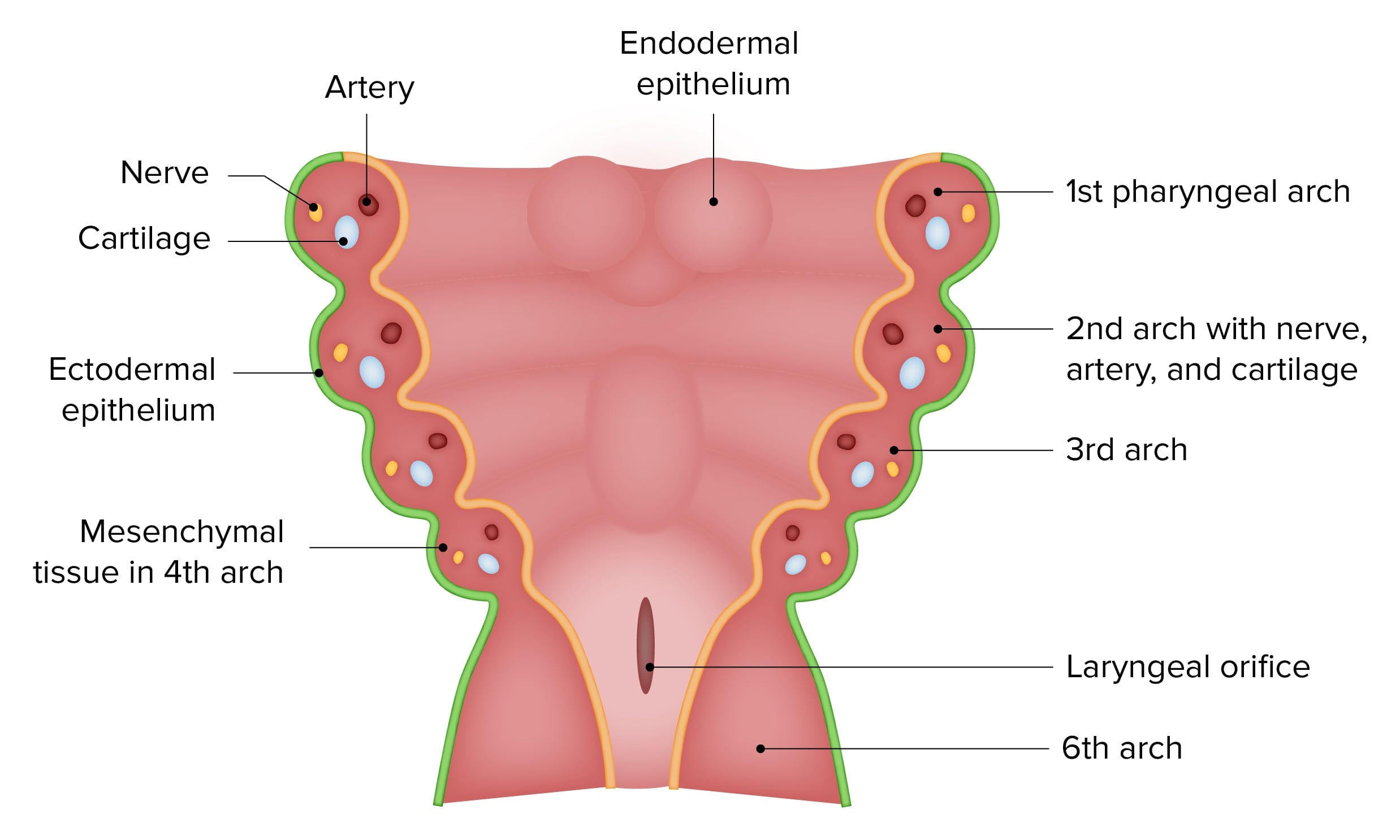
00:01 Now we're going to take a look at the regions that make up the pharynx. 00:08 The pharynx collectively is this space behind the nasal and oral cavities. 00:15 The choana is the aperture between the nasal cavity and the nasal pharynx. 00:21 And we see that it opens up right at the same time the hard palate becomes the soft palate. 00:27 So this area posterior to the choana is called the nasopharynx. 00:33 And it's innervated by the maxillary division of trigeminal, cranial nerve V2. 00:41 Further down in the neck, we have the hyoid bone and that area between the soft palate and the hyoid but posterior to the oral cavity is the oropharynx. 00:50 And that's innervated by the glossopharyngeal nerve or cranial nerve IX. 00:56 Further down, we see the esophagus. 01:00 And between the oropharynx and the esophagus is the last part of the pharynx that we call the laryngealpharynx or hypopharynx. 01:09 And that's going to be innervated by the vagus nerve or cranial nerve X. 01:18 So let's take a closer look at the nasopharynx. 01:23 Here we see the roof of the nasal pharynx formed by the basosphenoid and basiocciput areas. 01:31 And then posteriorly, we have the mucosal layer, followed by something called the pharyngobasilar fascia, a thick sheet of connective tissue here. 01:43 Then we have our pharyngeal muscles such as the superior constrictor muscle, and then we get into the pre-vertebral musculature as we move further posterior. 01:54 Eventually, we're going to hit the cervical vertebra and here we can see the arch of the atlas or C1. 02:02 The nasopharynx is where we also find a type of tonsil called the pharyngeal tonsil also called an adenoid. 02:10 The floor of the nasal pharynx is composed of the soft palate, the portion just posterior to the hard palate. 02:18 And this narrowing called the pharyngotympanic isthmus. 02:23 Anteriorly, we're bounded by the choana, and then laterally we see this lateral wall here where we find the opening of the pharyngotympanic tube, more commonly called the Eustachian tube. 02:37 And it's pharyngotympanic because it's the tube that conveys stuff from the pharynx to the tympanic area. 02:45 Tympanic refers to the tympanic membrane or the eardrum. 02:50 So, that's a way to remember that this tube is connecting to the middle ear cavity. 02:58 Beyond this, we have the oropharynx. 03:02 Again, limited by the hard palate above and the hyoid bone below, we have these two imaginary lines as our superior and inferior borders of the oropharynx. 03:15 Here we see the oropharyngeal isthmus. 03:18 Anterior to which we have the oral cavity. 03:21 Inferiorly, we see we have the base of the tongue, where we find the lingual tonsils, and then that little gap or tiny valley or vallecula between the base of the tongue and the epiglottis. 03:37 Posteriorly, we have more of those pharyngeal constrictor muscles. 03:41 And then here we see the lateral wall. 03:44 And we have these bumps or arches formed by the muscles beyond the mucosa here, such as the palatoglosall arch in the area of the palatoglossus, and the palatopharyngeal arch in the area of the palatopharyngeus. 03:57 And in between is where we find the tonsillar fossa for the palatine tonsil. 04:06 And this area here superiorly is where we transition from the nasal pharynx into the oropharynx. 04:13 Inferiorly, this is where we're going to have an opening into the laryngeal pharynx or hypopharynx. 04:20 And there's this little ridge here as serving as a landmark called passavant ridge. 04:29 Here we have the laryngopharynx starting around the area of the epiglottis which is the protector of the larynx. 04:39 And this keeps food from entering the larynx and into the trachea. 04:43 And this way we only have air going down into the lungs. 04:47 Instead, the epiglottis will flop down during swallowing and force food or liquids posteriorly into the esophagus. 04:57 The area between the epiglottis and the esophagus is that hypopharynx or laryngopharynx. 05:06 Here's a posterior view, where we can see the oral cavity, we're looking at it from behind. 05:12 So anteriorly is where we would find the oral cavity from here. 05:16 And we find the epiglottis protecting the laryngeal inlet or the opening for the airway. 05:24 And we would feel some cartilage in this area in the posterior aspect, we would feel the cricoid cartilage. 05:31 And on either side of that laryngeal cartilage, we would have a little space called the pyriform recess.
About the Lecture
The lecture Pharynx by Darren Salmi, MD, MS is from the course Upper Aerodigestive Tract.
Included Quiz Questions
Which nerve innervates the nasopharynx?
- CN V2
- CN V3
- CN V1
- CN X
- CN VII
Which nerve innervates the hypopharynx?
- CN X
- CN IX
- CN VII
- CN V2
- CN V3
Which layer of the posterior wall of the pharynx is the most posterior?
- Prevertebral musculature
- Superior constrictor
- Mucosal layer
- Pharyngobasilar fascia
- Sphenoid process
What best describes the location of the Eustachian tube?
- Lateral wall of the nasopharynx
- Medial wall of the nasopharynx
- Medial wall of the oropharynx
- Lateral wall of the oropharynx
- Lateral wall of the hypopharynx
Author of lecture Pharynx

Darren Salmi, MD, MS
Customer reviews
5,0 of 5 stars
| 5 Stars |
|
5 |
| 4 Stars |
|
0 |
| 3 Stars |
|
0 |
| 2 Stars |
|
0 |
| 1 Star |
|
0 |
