Playlist
Show Playlist
Hide Playlist
Pharyngeal Arches

-
Slides 04-16 Pharyngeal Arches.pdf
-
Reference List Embryology.pdf
-
Download Lecture Overview
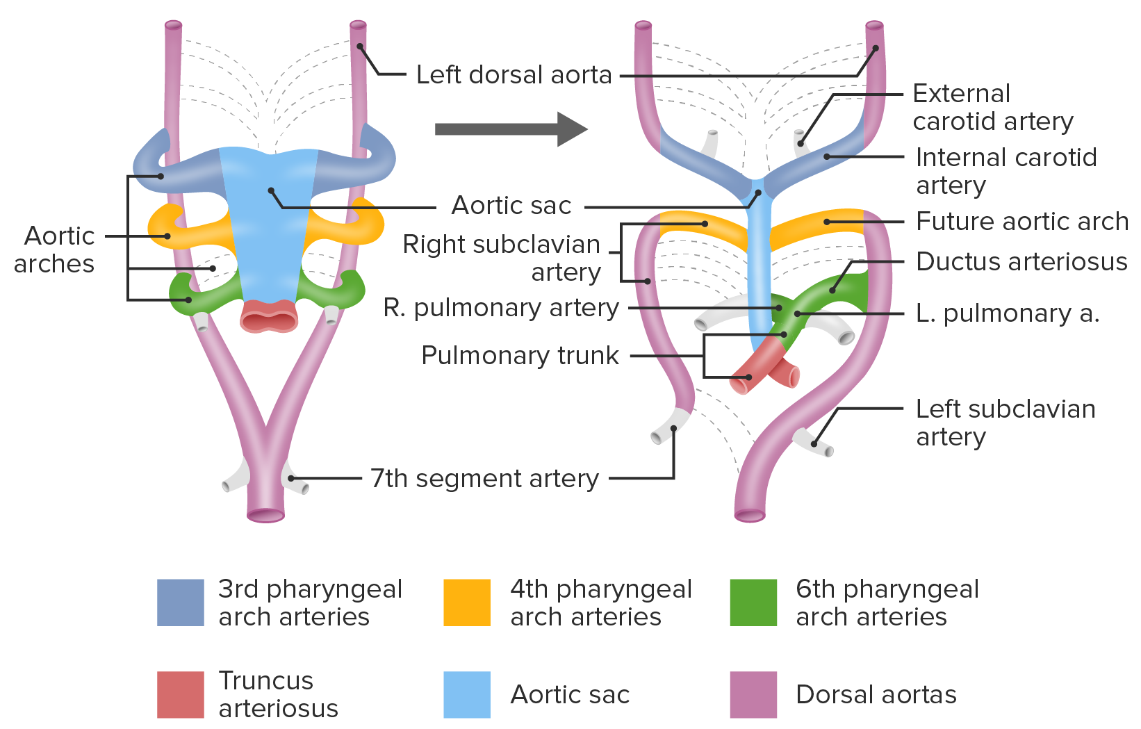
00:01 Hello. And now, we´re gonna discuss the pharyngeal arches. 00:05 Now, these go by a variety of names. 00:07 Pharyngeal arches, gill arches, branchial arches, but they´re all referring to the same thing which is a series of mesenchymal folds that develop along the neck during early, early development. 00:20 Before we have a face, we start to get these pharyngeal arches. 00:23 Now, they´re basically a little curving arch with the hanging loose side pointing inferiorly that´s gonna be full of mesenchyme. 00:33 And that mesenchyme is gonna develop into a variety of different structures. 00:36 As development proceeds from anterior to posterior, we develop five sets of arches. 00:44 So the first arch will be located anterior to the second, to the third, to the fourth, and so on. 00:51 Now, these arches are gonna form a variety of structures including most of our lower face. 00:56 They´re gonna be part of our ears, our throat, and along with that, several glands that contribute to our endocrine system and especially, to our calcium regulation. 01:07 So each pharyngeal arch contains a core that´s gonna have dense mesenchyme within it that´s going to become a cartilage rod which will then later, transition into bone, so will undergo endochondral ossification and that core of cartilage is associated with a muscle, and that muscle is associated with a nerve, and there´s also gonna be an artery supplying it. 01:31 Just a quick preview of heart development will tell you that the artery within each pharyngeal arch is called an aortic arch and they´re present here and they´re also gonna be contributing to most of our vasculature in the neck and head. 01:45 So that´s a brief preview of coming attractions. 01:48 Now, the first pharyngeal arch, the one that´s closest to the front is gonna become part of the incus and malleus inside of our ear. 01:58 So incus and malleus, the first two ossicles as well as our mandible, our jaw, and parts of the maxilla. 02:06 So the zygomatic bone, maxilla, are gonna be coming from the first arch. 02:10 Going a little further inferiorly, we have the second arch. 02:14 It´s going to form the last of the three ossicles so the middle ear bones, the stapes, the styloid process of the skull and part of the hyoid bone, specifically, its lesser horn are all coming from the second arch. 02:28 The third arch is gonna create the body and greater horn of the hyoid bone. 02:32 And the fourth and sixth arches create laryngeal cartilages. 02:36 Now, if that´s a bit abstract for you, I´ve got some good news. 02:40 This picture should make it clear that we´re laying down these arches from superior to inferior with the first arch contributing the malleus, the incus, parts of the mandible. 02:52 The second arch, the stapes, the styloid process, and the lesser horn of the hyoid bone. 03:00 The rest of the hyoid bone is coming from the third arch. 03:03 And finally, the fourth and sixth arches do not ossify but they remain cartilaginous and create the laryngeal cartilages. 03:11 Now, if you look to the left, you´ll see that a variety of cranial nerves are also growing into the pharyngeal arches. 03:18 In this case, cranial nerves 5, 7, 9, and 10. 03:23 And right now, it´s a good idea just to drill those numbers 5, 7, 9, 10, because they´re gonna be very important as this development proceeds. 03:32 Now, in the head and neck, we have structures called somitomeres and they´re very much like somites that we´ve seen earlier and remember that somites were distinct little beads developing on either side of the neural tube that were innervated by a nerve and then, drag that nerve behind it as it would migrate to become different muscles in the body. 03:51 Somitomeres are very similar except they don´t form distinct bulges like the somites and we´re gonna have somitomeres migrate into each arch and as they do so, they´re gonna drag their nerve supply behind them. 04:02 That nerve supply is coming from cranial nerves 5, 7, 9, and 10. 04:08 So as they move into the arch, they´re going to associate with the various cartilaginous and bony structures that are developing there. 04:15 And that cranial nerve will thereafter not just innervate those muscles but move whatever cartilaginous and bony structures those muscles are attached to. 04:24 Now, the first arch, the somitomeres that migrate into it are going to form the muscles of mastication. 04:31 AKA the muscles that we use to eat. 04:34 Now, by necessity, those muscles have to attach to the jaw. 04:38 So muscles of mastication as well as part of our digastric muscle and our tensor tympani muscle all come from the first arch. 04:47 Now, the first arch gave rise to the incus, the malleus, the mandible, as well as some of the facial bones. 04:55 So it makes sense that the muscles that are going to be attaching to those bones are coming from the first arch and the main thing to remember about their innervation is that they´re all innervated by V3, the mandibular branch of the trigeminal nerve and anytime that nerve has to be dragged behind those muscles as it moves, it´s going to leave a course through the bone. 05:16 Next up, we have the second pharyngeal arch. 05:20 The somitomeres that migrate in here are pulling behind them the branches of the facial nerve. 05:26 So the facial nerve innervates all of these somitomeres and they then, migrate to any bones that are gonna be associated with the second arch. 05:35 So the styloid process, part of the hyoid bone and the stapes. 05:40 That tiny little stapedius muscle inside the middle ear is innervated by the facial nerve because the stapes comes from the second arch. 05:48 Now, much more prominently, the muscles of facial expression that migrate from their starting point about here all over the face are gonna pull the facial nerve behind them and that´s why the facial nerve has that extended branching pattern on the face as it moves around and is dragged as the muscles migrate. 06:09 So as the muscles migrate across the face, the facial nerve is passively pulled behind. 06:14 Now, an interesting exception to this rule is the third arch. 06:19 It´s not an exception in that it does anything that different but it creates very little muscle. 06:25 In fact, the somitomeres that go into the third arch eventually create a single muscle called the stylopharyngeus and that muscle is a longitudinal muscle inside the pharynx. 06:36 It´s innervated by the glossopharyngeal nerve and the glossopharyngeal nerve innervates one and only one somitomere-derived muscle and that´s it. 06:45 Now, as opposed to the third arch, the fourth arch gives rise to a great many muscles. 06:51 All the muscles of the palate and the pharynx except for the stylopharyngeus are all derived from the fourth arch and the fourth arch is innervated by the vagus nerve. 07:02 So when you associate motor activity of the vagus nerve with anything, it´s probably the palate, the pharynx, and the larynx. 07:09 Now, let´s move on to the sixth pharyngeal arch. 07:14 The somitomeres that enter the sixth arch are also innervated by the vagus nerve but they´re specifically innervated by its recurrent branch. 07:23 A very separate and distinct branch of the vagus nerve goes into the sixth arch, innervates the muscles that are found there and these muscles will become all the muscles of the larynx, the ones that allow us to speak. 07:36 So all of our muscles of phonation are coming from the sixth arch and are innervated by the recurrent branch of the laryngeal nerve or the current laryngeal nerve which is a branch of the vagus nerve. 07:48 The only exception is the one from the fourth arch, the cricothyroid muscle. 07:55 Now, you may be thinking to yourself that I made an error and I didn´t talk about the fifth arch. 08:01 The fact is these structures were described first in fishes and gilled animals and there were in fact one, two, three four, five, six, sometimes seven gilled structures inside these fishes. 08:14 When it came time to organize human embryology and figure out what was going on, they found that the fifth arch structures that exist in some animals have no homologs in humans. 08:25 That´s why humans have one, two, three, four, no five, and a sixth pharyngeal arch. 08:31 Now, let´s talk about things that can go wrong with the first arch. 08:35 There are a variety of problems. 08:36 Treacher-Collins syndrome, Pierre-Robin sequence, and Agnathia. 08:41 Now, Treacher-Collins syndrome results from too few neural crest cells migrating into the first arch and too little tissue there to actually build things. 08:52 So we have underdevelopment of the jaw. 08:54 That´s called micrognathia. 08:56 You´re gonna have deformity of the zygomatic bones, part of the bony structures that come from the first arch in the cheek as well as the ossicles and external ears. 09:05 So difficulty hearing is another hallmark of Treacher-Collins syndrome. 09:09 And mutations are resulting in too few neural crest cells migrating here in the first place. 09:16 So it´s a genetic issue that´s brought about with too few neural crest cells making it to the first arch in the first place. 09:23 Something that may initially appear similar but is very distinct is Pierre-Robin sequence and a sequence implies one thing goes wrong and sets off a chain of effect thereafter. 09:35 In the case of Pierre-Robin sequence, you´ve got a small jaw. 09:39 And that small jaw can occur for reasons that we don´t quite understand. 09:43 Most prevalent theory right now is that its intrauterine positioning and the jaw is pressed against the uterus or against some other structure and is unable to develop. 09:53 And because the jaw can´t develop anteriorly, we wind up with too small a jaw, micrognathia but it also means that the tongue is too far posterior and that does not allow the palate to close, so we have a cleft palate. 10:08 So the Pierre-Robin sequence is micrognathia, posterior tongue, and cleft palate. 10:14 Last and quite possibly less common than anything else, we have agnathia. 10:21 Failure of the jaw to form in the first place. 10:24 This is a severe problem related to migration of neural crest cells into the first arch and flat out failure of the first arch to form normally. 10:33 Thankfully, this is a very uncommon thing to encounter but it does have a name and it does occur to some degree. 10:40 So be on the watch for it if you´re ever on pediatric or OB-GYN rotations and looking for something very strange on ultrasound. 10:48 Alright, thank you very much.
About the Lecture
The lecture Pharyngeal Arches by Peter Ward, PhD is from the course Development of the Nervous System, Head, and Neck. It contains the following chapters:
- Introduction to the Pharyngeal Arches
- The Different Arches
- Characteristics of First Arch Syndromes
Included Quiz Questions
Which pharyngeal arch will give rise to the mandible?
- First
- Second
- Third
- Fourth
- Sixth
Which pharyngeal arch will give rise to the last of the three ossicles?
- Second
- First
- Third
- Fourth
- Sixth
The fourth and sixth pharyngeal arches will give rise to which of the following structures?
- Laryngeal cartilages
- Maxilla
- Hyoid
- Styloid process
- Malleus
What is the motor nerve supply to the first pharyngeal arch?
- CN V3
- CN V1
- CN V2
- CN VII
- CN IX
What is the only muscle that is derived from the third pharyngeal arch?
- Stylopharyngeus muscle
- Digastric muscle
- Stylohyoid muscle
- Lateral pterygoid muscle
- Tensor veli palatini muscle
Which of the following describes the condition in which the mandible is absent?
- Agnathia
- Pierre-Robin sequence
- Second Arch syndrome
- Treacher-Collins syndrome
- Potter sequence
Author of lecture Pharyngeal Arches

Peter Ward, PhD
Customer reviews
5,0 of 5 stars
| 5 Stars |
|
3 |
| 4 Stars |
|
0 |
| 3 Stars |
|
0 |
| 2 Stars |
|
0 |
| 1 Star |
|
0 |
Elegí esta calificación porque explicó de forma sensilla de entener y recordar lo básico del desarrollo y patologias de los arcos faríngeos, ahora queda ib¿vestigar más sobre dicha información. Me gustó que pone la variedad y cantidad de imagenes que proyecta justo en el momento exacto que explica, eso hace más entendible la conferencia. Recomendaría este vídeos a mis compañeros de facultad.
Just AMAZING!!!! Thank you Dr Peter for making this lecture so easy. This made me understand more rather than mugging up the derivatives.
Lecturio should open their own medical school in the Caribbean and people could actually learn down there ! keep it up !
