Playlist
Show Playlist
Hide Playlist
Pericyte

-
Slides 02 Human Organ Systems Meyer.pdf
-
Reference List Histology.pdf
-
Download Lecture Overview
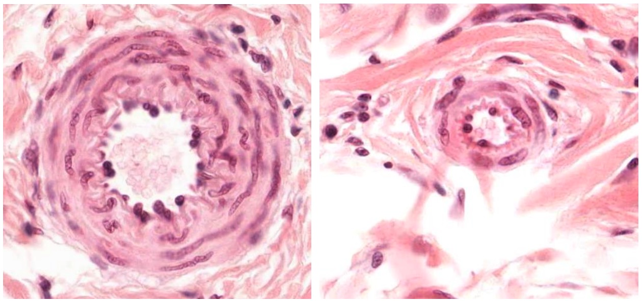
00:00 Here, in this section, you can see a repeat of those small little arteries, we call an arteriole. They are very very small. You’re looking here at a very high magnification. But still, you can point out the nuclear eye bulging of the endothelial cell, the endothelium. You can even make out a very small component, wiggly line there, which is the internal elastic lamina. But notice, there are only one or two layers of smooth muscle around each of these vessels. And then, you really can’t see components of the adventitia on the outside. It just blends in with surrounding connective tissue. But I want to mention the pericyte. On the right-hand side, you can see a little tiny vessel. 00:58 This little tiny vessel is surrounded by an endothelium. Remember, the endothelium is the lining of the capillary. If I just take away the label, have a look at this tiny little vessel. 01:13 Try and pick out a nucleus. It will be the endothelial cell nucleus. I know this is very hard, and it is very hard. But just on the outside of this very, very small vessel, it’s a little capillary actually, is another nucleus we call the nucleus of the pericyte. This pericyte is very important. It wraps around very small vessels, particularly capillaries, and it has a number of functions. You know, during my early research career, I used to study pericytes and the function they had, because at that stage, they are thought to inhibit the growth of capillaries. And therefore, maintain a certain proportion of a tissue that’s occupied by the blood circulation. And one of the big problems with cancers is that once the cancer cells spread and get into other organs, they attract the blood supply. And therefore, more and more blood vessels grow into the tumor, and therefore, the surrounding healthy tissue is starved and dies. Well, my interest was on these pericyte cells because that was said to be inhibitory to maintain low growth of blood capillaries. And I thought if you could actually try to get those pericytes to stop blood vessel growth in tumors, then you’d be able to limit the growth of the tumor and the blood flow through them. 02:45 And that’s led on to further research in trying to control what we call angiogenesis, the generation of new blood vessels, particularly, in relation to controlling tumor growth and spreading of cancers. Well, these pericytes also have other functions. They said to be able to divide and actually turn into maybe smooth muscle cells or adventitia cells, again, in situations where the capillary bed may be expanding and growing further. They are actually surrounded by the external lamina of the endothelial cell or a term that we call the basal lamina because these are, in fact, endothelium. And because the pericyte is surrounded by the basal lamina of the endothelium or shares the basal lamina of the endothelium, it’s actually not a connective tissue cell. Sometimes we often refer to a pericyte as being a connective tissue cell. But because they are separated from the connective tissue elements by this basal lamina means that they’re really epithelial. I’m sure in the future these pericytes will be assigned a lot more functions and a lot more importance.
About the Lecture
The lecture Pericyte by Geoffrey Meyer, PhD is from the course Cardiovascular Histology.
Included Quiz Questions
Which of the following statements regarding pericytes is NOT correct?
- They are cancerous cells.
- They are epithelial cells.
- They can turn into smooth muscle cells.
- They may have inhibitory effects on angiogenesis.
Which of the following is MOST ACCURATE regarding the histology of arterioles and arteries?
- The smooth muscle layer is less pronounced in arterioles.
- The external elastic lamina is absent in arteries.
- The internal elastic lamina is absent in arterioles.
- The tunica intima is very thick in arterioles.
- The are no major differences in the histology of arteries and arterioles.
What separates the pericytes from the surrounding tissue?
- Basement membrane
- Reticular lamina
- Gap junctions
- Tight junctions
- Desmosomes
Author of lecture Pericyte

Geoffrey Meyer, PhD
Customer reviews
5,0 of 5 stars
| 5 Stars |
|
5 |
| 4 Stars |
|
0 |
| 3 Stars |
|
0 |
| 2 Stars |
|
0 |
| 1 Star |
|
0 |
