Playlist
Show Playlist
Hide Playlist
Patient Introduction and Review of Neck and Back Anatomy

-
Slides Physical Exam Neck and Back Pain Introduction.pdf
-
Reference List Physical Examination.pdf
-
Download Lecture Overview
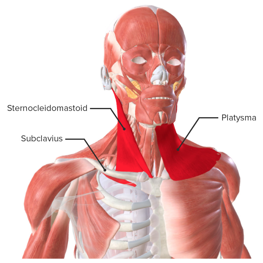
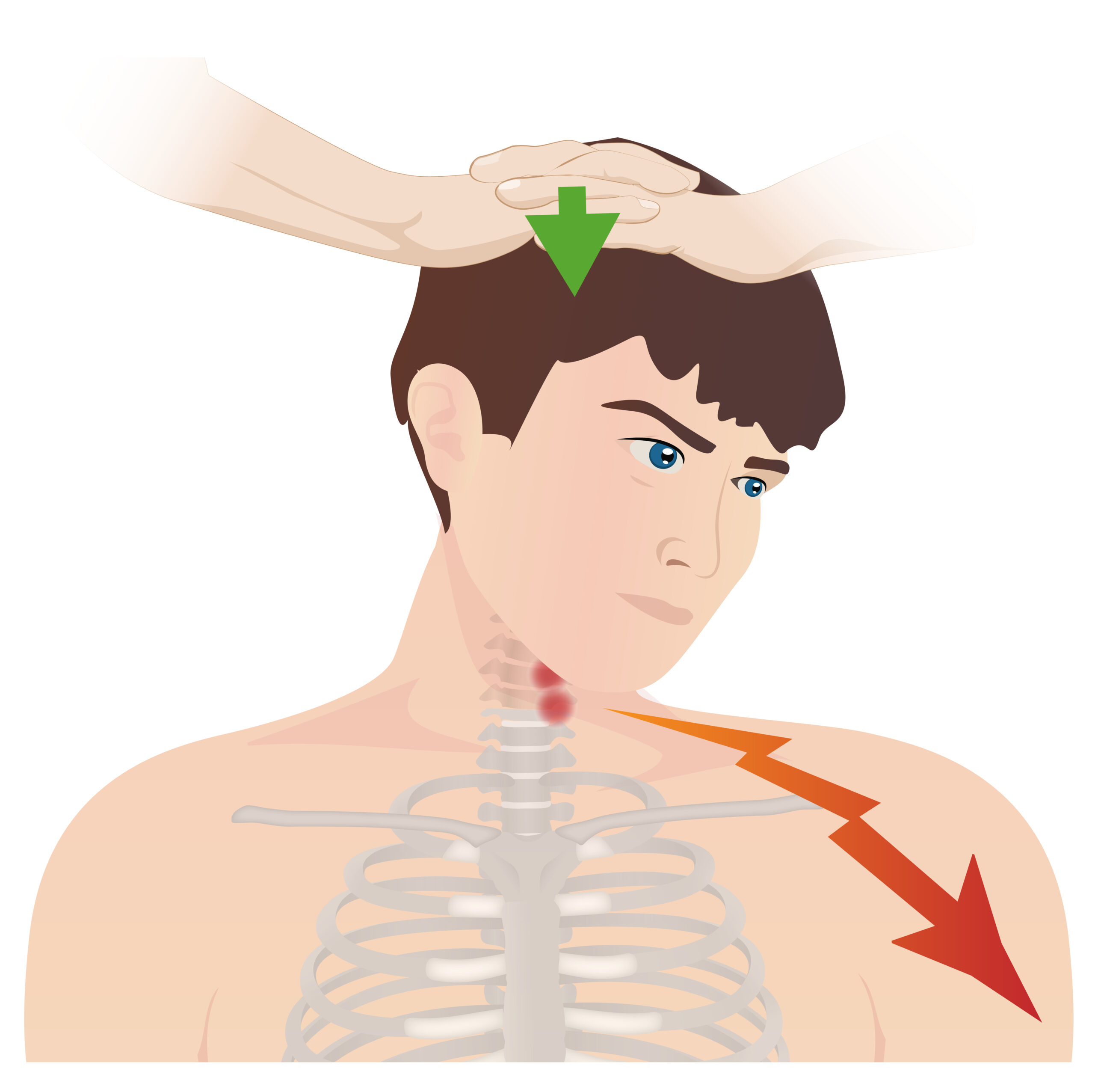
00:01 Okay, let's start with the case of a patient with back pain. 00:05 A 56-year-old man presents with low back pain that has been worsening for the last year. 00:10 He describes it as a sharp twinge that often radiates down his left leg. 00:15 Now, there's a lot of potential possibilities for this person's pain and it's going to be really important for us to use our exams skills to tease apart what's going on. 00:23 First off, whenever a patient has low back pain, I like to divide it into three broad categories of potential disease. 00:30 Perhaps the most common is just a simple muscle strain. Next up is spondylosis. 00:36 This is a broad category that simply means disease of the spine. 00:40 There could be just axial discomfort which we'll talk more about, radiculopathy, involving the nerve roots; myelopathy, encroachment on the spinal cord itself, and often times a mixture of all of the above. 00:53 And then lastly, what we'll focus less on is the miscellaneous category, you know, cancers, rheumatologic etiologies, infectious causes like discitis or osteomyelitis, and of course, peripheral neuropathy simply causing pain referring to that area. 01:09 I've mentioned elsewhere in the introductory lecture, the importance of the physical exam, vis á vis, radiologic imaging and this is a great example. 01:20 There was a study that looked at 100 asymptomatic persons over the age of 40 who had a cervical neck MRI. 01:27 It turned out that 28 of them had evidence of herniated discs and or spinal stenosis. 01:33 These are folks with no symptoms whatsoever. 01:35 So if we just image everybody with back pain, we're going to find disc bulges here, facet joint arthropathy there, maybe even spinal stenosis in some areas; but if those areas don't directly correlate with the findings on our physical exam, then we shouldn't go and perform surgery on those things because it can be, you're going to be causing more problems than you started off with. 01:58 This is why it's so important to correlate our physical exam with radiologic imaging. 02:02 With that, lets look at the potential muscles involved in low back pain. 02:08 We always talk about the muscles that are immediately paraspinal and then some that are further, wrapping around the sides. 02:15 Often times, patients with mid thoracic pain in the back have issues with the rhomboid musculature. 02:22 People with posterior shoulder pain rarely have a problem with the shoulder itself, it is more to do with the paraspinal musculature on either side. 02:33 Other relevant and anatomy to recall is that the spinal column is broken up into four sections: cervical, thoracic spine, and lumbar spine and then the bottom is just the sacrum which we're not highlighting in this image. 02:46 Each section of the spine has it's owns unique properties that allow for the structure and function to dictate movement. 02:53 For example the cervical spine is the most flexible part of your spine. 02:57 It allows you to laterally flex, forward flex, extension, rotation, etcetera, whereas the lumbar spine tends to have somewhat more limited range of motion. 03:05 I'll draw your attention on this slide to highlight the intervertebral discs which are these rather cartilaginous structures that occur between each vertebra, and those intervertebral discs can have specific diseases of their own. 03:19 Inside an intervertebral disc is the nucleus pulposus which can herniated out and encroach upon the spinal canal. 03:26 This is lumbar vertebrate, which again, also have intervertebral discs. 03:32 The structure here, again, allows for somewhat less movement than the cervical bodies. 03:37 And also remember that ones you get down to around L3 or L2, you no longer have a spinal cord instead you just have the cauda equina, the horse tail of reticular nerves before they exit the spinal canal. 03:51 A quick view of just one single vertebral body. 03:57 Remember that on top of the body shown in this image would be an intervertebral disc. 04:02 Also, you'll note that there are facets. 04:05 The facets are the joints, the articulations between one vertebral body and the vertebral body above it and below it, and while we don't typically think about arthritis of the vertebral bodies, the arthritis is occurring at the facets themselves and we'll look more closely at that in the next few slides. 04:25 In the center is the vertebral foramen also-called the spinal canal and take note of the spinous process, that's the most prominent palpable part of the vertebral body on the back of the spine. 04:36 Looking at a lumbar vertebra. 04:41 Take note again of those facets that are articulating between one vertebral body and the next, the superior articular facet above and the inferior articular facet below.
About the Lecture
The lecture Patient Introduction and Review of Neck and Back Anatomy by Stephen Holt, MD, MS is from the course Examinations of the Neck and Back Region.
Included Quiz Questions
Which part of a vertebral body is palpable on physical examination?
- Spinous process
- Superior articular facet
- Transverse facet
- Lamina
- Transverse process
Author of lecture Patient Introduction and Review of Neck and Back Anatomy

Stephen Holt, MD, MS
Customer reviews
5,0 of 5 stars
| 5 Stars |
|
5 |
| 4 Stars |
|
0 |
| 3 Stars |
|
0 |
| 2 Stars |
|
0 |
| 1 Star |
|
0 |
