Playlist
Show Playlist
Hide Playlist
Parietal Bone

-
Slides Anatomy Skull.pdf
-
Download Lecture Overview
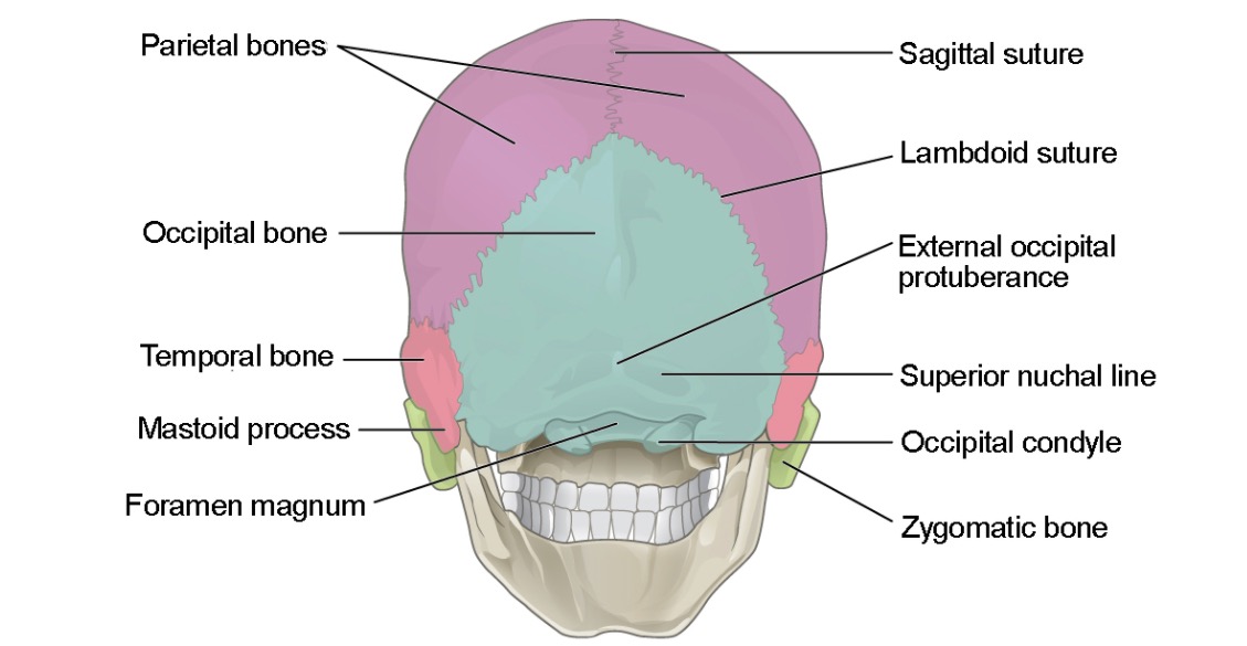
00:01 Now let's talk about the parietal bones. 00:03 The parietal bones are paired quadrilateral bones that make up the majority of the cranial roof or the calvaria, as well as its lateral aspect. 00:13 When we discuss the parietal bone, we have to touch upon several points. 00:17 One, the various topographical markings located on its external and internal surfaces and two, its four borders and angles. 00:26 So let's first begin our discussion on the topographical markings of the parietal bone. 00:32 Externally on the parietal bone, a continuation of the curves superior and inferior temporal lines of the frontal bone can be seen. 00:41 Once again, these lines mark the superior border of the temporal fossa. 00:45 On the internal surface of the temporal bone, there are many markings made up by the impressions of the cerebral gyri and the meningeal vessels, but the most important landmark is the groove for the superior sagittal sinus. 01:00 This groove is formed by the union of the two parietal bones along the sagittal suture. 01:06 Now let's move our discussion along to the borders of the parietal bone. 01:11 As I mentioned in the introductory slide, the parietal bone has four borders. 01:16 By borders we mean edges at which the parietal bone articulates with the surrounding cranial bones. 01:23 These four borders include the frontal border, where it articulates with the frontal bone, the sagittal border where it meets the opposite parietal bone, the inferior squamosal border where it meets the temporal bone and the greater wing of the sphenoid, and we have the occipital border where it meets the occipital bone. 01:48 And now for the last remaining portion in our discussion of the parietal bone, its angles. 01:54 The angles of the parietal bone, conveniently also numbered four, and they are the frontal anterosuperior angle that's located at the bregma, which marks the intersection of the coronal and sagittal sutures. 02:09 Additionally, this angle also marks the location of the anterior fontanelle in neonates. 02:16 Also on this image we have the sphenoidal anteroinferior angle, which is located at the pterion. 02:23 This is a craniometric point which marks the intersections of the frontal, parietal sphenoid, and the temporal bones. 02:31 Additionally, at this angle, but on the anterior surface, lies a groove for the frontal branches of the middle meningeal arteries. 02:41 The third angle is the occipital posterosuperior angle. 02:45 This lies at the cranial metric point called the lambda and this marks the intersection of the sagittal lambdoid sutures of the parietal and occipital bones respectively. 02:56 Plus more, this angle marks the location of the posterior fontanelle in neonates. 03:02 And then lastly, we have the mastoidal posteroinferior angle that lies at the craniometric point known as the asterion and this marks the intersection of the occipital, parietal and the mastoid portion of the temporal bones. 03:18 Additionally, the interior surface of this angle is located at which the transverse and the sigmoid sinuses meet. 03:28 With this, we conclude our discussion of the parietal bone. 03:32 Next we move on to the ethmoid bone, which is the last portion of the neural cranium we will be discussing in this presentation.
About the Lecture
The lecture Parietal Bone by Craig Canby, PhD is from the course Head and Neck Anatomy with Dr. Canby.
Included Quiz Questions
Which point marks the location of the intersection of the transverse and sigmoid sinuses?
- Mastoidal posteroinferior angle
- Occipital posteroinferior angle
- Occipital posterosuperior angle
- Frontal anterosuperior angle
- Mastoidal posterosuperior angle
Where does the superior sagittal sinus lie with respect to the parietal bones?
- At the union of the parietal bones
- Anterior to the union of the parietal bones
- Posterior to the union of the parietal bones
- Lateral to the parietal bones
- The superior sagittal sinus runs perpendicular to the union of the parietal bones.
Author of lecture Parietal Bone

Craig Canby, PhD
Customer reviews
5,0 of 5 stars
| 5 Stars |
|
5 |
| 4 Stars |
|
0 |
| 3 Stars |
|
0 |
| 2 Stars |
|
0 |
| 1 Star |
|
0 |
