Playlist
Show Playlist
Hide Playlist
Parasympathetic Nervous System (PSNS)

-
Slides 1 NervousSystem BrainAndNervousSystem.pdf
-
Reference List Anatomy.pdf
-
Download Lecture Overview
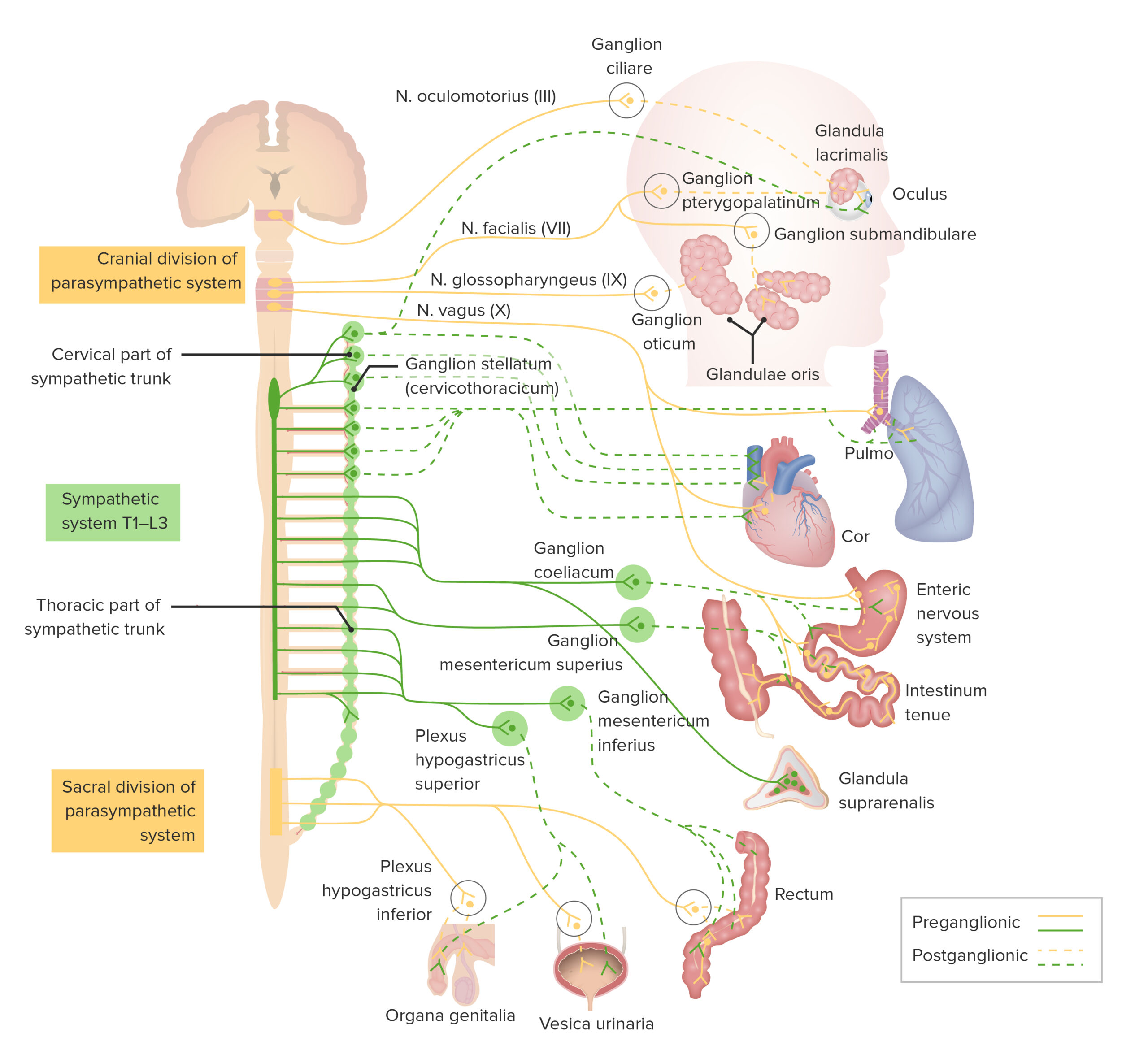
00:01 Now, I want you to understand in greater detail the parasympathetic system. Another term for this system is the craniosacral system because the cell bodies or the preganglionic neurons reside in certain cranial nerve nuclei and in the terminal portion of the spinal cord. 00:27 So, one of the cranial nerve nucleus that contains parasympathetic fibers is cranial nerve III. 00:39 Cranial nerve III is shown in through here. This is the oculomotor nucleus. Then its parasympathetic fibers can be seen here extending outward to the ganglion here. 00:54 We’ll talk more about these ganglia. Another point of origin of parasympathetics from cranial nerve nuclei is that of the facial nerve, cranial nerve VII that is shown here. Then you can see it extending out toward the periphery. It will synapse with two ganglia that we see here. 01:15 We’ll identify those as well shortly. Cranial nerve nucleus IX, the glossopharyngeal nucleus is shown in through here. You can see the glossopharyngeal nerve extending outwards conveying the parasympathetic fibers that synapse with the ganglion. Then the last cranial nerve that conveys parasympathetic nerve fibers will be cranial nerve X. That’s the vagus nerve. 01:41 Here is its nucleus. Here is the peripheral distribution, very, very wide spread as you can appreciate. Lastly, pelvic splanchnic nerves from sacral segments S2, S3, S4. The nerve cell bodies of these preganglionic nerve fibers reside here. Then those nerve fibers will extend outwards toward the periphery and then synapse with ganglia in the walls of the organs that they innervate. Now that you know where the preganglionic nerve cell body resides for the parasympathetic system, what ganglion does each one of those components synapse with? So let’s take a look at that. We’ll begin with cranial nerve III, the oculomotor nucleus. Its nerve cell body will extend an axon outwards until it synapses with the ganglion that we see here and that is the ciliary ganglion. Cranial nerve VII, the facial nucleus, its preganglionic axons are going to synapse with two ganglia, the pterygopalatine ganglion here and then the submandibular ganglion that we see below. Cranial nerve IX, the glossopharyngeal nerve, it will extend its preganglionic axon outwards and synapse with the otic ganglia. Then from here, it will send its postganglionic nerve axon or fiber to the parotid gland. The mnemonic here, GOP or acronym is glossopharyngeal otic ganglion and parotid gland. Cranial nerve X extends outwards to innervate the viscera of the thorax, mid-gut and the very proximal portions of the hindgut. The ganglia are in the wall of those particular structures. Lastly, pelvic splanchnic nerves which originate from S2, S3, S4, those sacral spinal cord segments, those preganglionic fibers extend outwards and will synapse in the terminal ganglia of the distal hind gut as well as the pelvic viscera.
About the Lecture
The lecture Parasympathetic Nervous System (PSNS) by Craig Canby, PhD is from the course Autonomic Nervous System (ANS).
Included Quiz Questions
Which of the following statements regarding the parasympathetic division of the ANS is most accurate?
- Cell bodies of preganglionic neurons reside in the brain stem and the sacrum.
- Preganglionic fibers are short, and postganglionic fibers are long.
- Each preganglionic neuron gives rise to many postganglionic neurons.
- Ganglia are situated close to the spinal cord.
- It has a stimulatory effect on most of the organ systems.
Which of the following combinations of synapses is NOT correct?
- CN X—pterygopalatine ganglion
- CN III—ciliary ganglion
- CN VII—submandibular ganglion
- CN IX—otic ganglion
- Splanchnic nerves—terminal ganglia of the hindgut and pelvic viscera
Which of the following is the last cranial nerve to carry parasympathetic nerve fibers?
- Vagus nerve
- Facial nerve
- Splanchnic nerves
- Oculomotor nerve
- Glossopharyngeal nerve
Author of lecture Parasympathetic Nervous System (PSNS)

Craig Canby, PhD
Customer reviews
5,0 of 5 stars
| 5 Stars |
|
3 |
| 4 Stars |
|
0 |
| 3 Stars |
|
0 |
| 2 Stars |
|
0 |
| 1 Star |
|
0 |
I'm a first year medical student and the lectures that we have had on the autonomics have not been clear or understandable at all. The set of lecture materials and the lectures that Lecture has provided has GREATLY increased my understanding and made my exam prep much easier.
It contains the keypoints. Neuroanatomy is hard because it´s specific and has lots of terms. Dr. Craig is making them simple and understandable!
Excellent lecture with very clear points that are made and in excellent chronological order. A fantastic way to get a brief overview and a great way to engrain the topics touched upon in the article.
