Playlist
Show Playlist
Hide Playlist
Palmar Aspect – Anatomy of the Hand

-
Slides 07 UpperLimbAnatomy Pickering.pdf
-
Download Lecture Overview
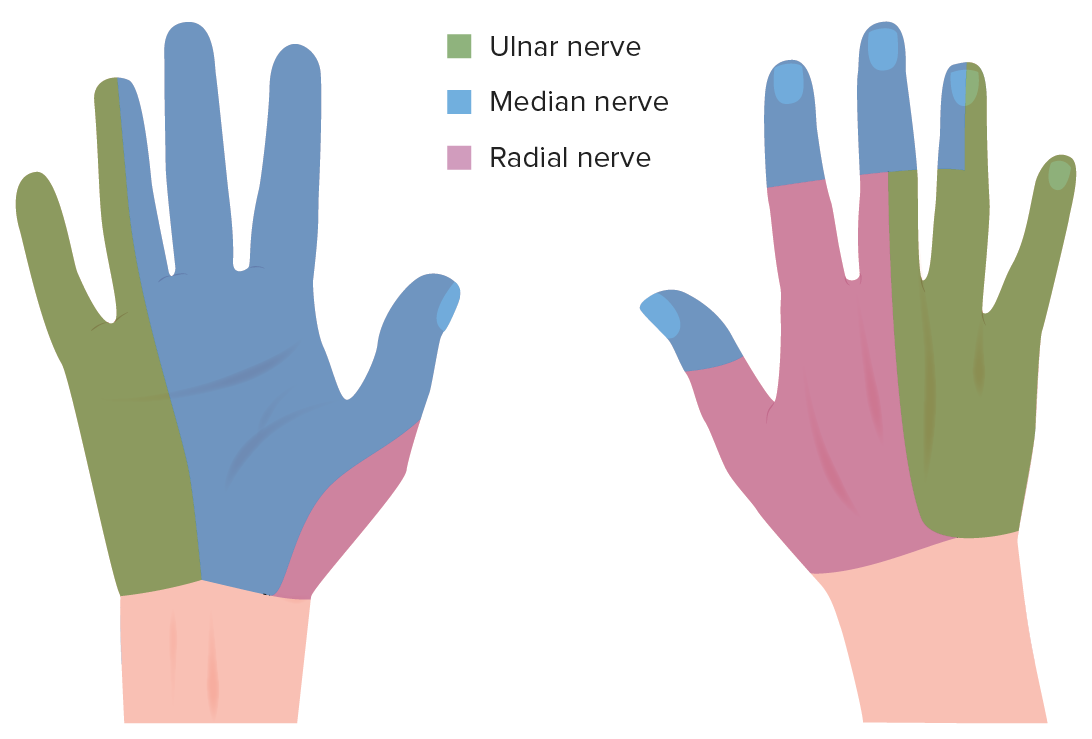
00:00 So now let’s move on to the palmar aspect of the hand. And the palmar aspect of the hand contains a whole series of intrinsic muscles that allow the hand to assume many different positions. And also, the tendons of the extrinsic muscles that originate in the forearm pass through the carpal tunnel to the hand. 00:23 Again on the slide, we can see we’ve got the thumb. We’ve got the second, third, fourth and fifth digit. So here we have the lateral aspect and here we have the medial aspect. 00:35 We can divide the palmar aspect of the hand into a number of compartments, and we can identify numerous muscles within those compartments. So here again on the slide, we have the picture. 00:48 We have this nice central compartment that contains mostly some deep muscles, but also the palmar aponeurosis. And the palmar aponeurosis is continuous here with the antebrachial fascia. 01:02 It also connected to a long muscle within the forearm, palmaris longus, and this helps to tighten this palmar aponeurosis. The palmar aponeurosis is important for protection. 01:14 It’s also important in being able to form the grip. It tightens the skin when you’re forming a grip. So, another space we can see is known as the hypothenar eminence. The hypothenar eminence is located medially within the hand, and we can see there’s a collection of muscles here, so the hypothenar eminence. Then we have the thenar eminence that is associated with the thumb. So we can see the thenar eminence here. We then have a small adductor compartment, which is just here, in between the thenar eminence and the central compartment. And then deep in between the metacarpals, we have an interosseous compartment. 01:57 And we are now going to explore all of these individual compartments. 02:02 Let’s have a look at the palmar fascia, first of all, and look at the palmar aponeurosis. 02:13 And we can see that the palmar aponeurosis is running over the whole palm but it is actually thin over the hypothenar and the thenar eminences. So it’s thin laterally, it’s thin medially. 02:28 But it thickens centrally and this is where we have this triangular-shaped palmar aponeurosis. 02:35 The apex of the palmar aponeurosis here is continuous with the palmaris longus muscle as I mentioned. And distally, it forms four longitudinal bands. And these longitudinal bands pass over the digits and support the digital sheaths that enclose the flexor tendons of flexor digitorum superficialis, FDS, and flexor digitorum profundus, FDP. 03:04 Remember, these muscles are originating in the forearm and they’re passing through the central compartment deep to palmar aponeurosis to pass through the digits. And these muscles are enclosed by the digital sheaths. And these longitudinal bands help to support those sheaths. 03:25 So let’s now look at the thenar eminence. This collection of muscles is associated with the thumb. We can see the thenar eminence is here. What we’ve done is we’ve removed the palmar aponeurosis on this diagram. So now we can just concentrate on this space here, the thenar eminence. The lateral aspect of the palm, the thenar eminence, contains three muscles: opponens pollicis, abductor pollicis brevis, and flexor pollicis brevis. 03:56 These muscles are primarily responsible for opposing the thumb, for opposition of the thumb, so it can assume a very unique position where you can draw the thumb across the palm of your hand, opposition. We can see these muscles here. Deep within the thenar eminence, we can see opponens pollicis. We can see this muscle here, opponens pollicis. 04:20 And then we can see that what were being cut are two muscles. We have flexor pollicis brevis here, flexor pollicis brevis. We can see the two cut-ends over here. So we have this muscle, flexor pollicis brevis, and we have a part of it here. And then more laterally, we have abductor pollicis brevis. So we can see abductor pollicis brevis here and we can see where it has been cut here. You also have a slightly deeper head of flexor pollicis brevis, and we can just make that out deep. It’s at the same plane as opponens pollicis. 04:53 So these are the three muscles that are within the thenar eminence. We can see the muscles here in this table highlighting their origins and insertions. And all three of these muscles come from the flexor retinaculum and specific tubercles of the scaphoid and trapezium bones. 05:12 So, all of these three muscles have a common origin. Opponens pollicis inserts onto the lateral side of the first metacarpal. Abductor pollicis brevis and flexor pollicis brevis attach to the lateral side of the proximal phalanx of the first digit. So just make sure you’re happy with the origins and insertions and refer back to the diagram if necessary. 05:40 All of these muscles, your thenar eminence, are supplied by a branch of the median nerve, and this is known as the recurrent branch of the median nerve. This nerve runs very superficial and can easily be damaged. But these three muscles are supplied by the recurrent branch of the median nerve. The function of these muscles really is as their name suggests. 06:06 So, opponens pollicis helps to oppose the thumb. Abductor pollicis brevis abducts the thumb and it also supports opposition. And flexor pollicis brevis helps to flex the thumb. 06:19 You should make sure you’re happy with the movements of the thumb.
About the Lecture
The lecture Palmar Aspect – Anatomy of the Hand by James Pickering, PhD is from the course Upper Limb Anatomy [Archive].
Included Quiz Questions
Which muscle within the forearm is connected to the palmar aponeurosis?
- Palmaris longus
- Extensor pollicis longus
- Abductor pollicis longus
- Extensor pollicis brevis
- Extensor indicis
Which statements regarding the thenar eminence are correct? Select all that apply.
- It is located laterally within the palm.
- It contains the opponens digiti minimi.
- It contains the opponens pollicis.
- It contains the abductor pollicis brevis.
- It contains the flexor pollicis brevis.
Which nerve supplies the muscles of the thenar eminence?
- Recurrent branch of the median nerve
- Anterior interosseous nerve
- Palmar cutaneous nerve
- Palmar digital branch of the median nerve
- Ulnar nerve
Author of lecture Palmar Aspect – Anatomy of the Hand

James Pickering, PhD
Customer reviews
5,0 of 5 stars
| 5 Stars |
|
5 |
| 4 Stars |
|
0 |
| 3 Stars |
|
0 |
| 2 Stars |
|
0 |
| 1 Star |
|
0 |
