Playlist
Show Playlist
Hide Playlist
Osteon

-
Slides 08 Types of Tissues Meyer.pdf
-
Reference List Histology.pdf
-
Download Lecture Overview
00:01 Well I mentioned earlier that the osteon is the functional and structural unit of the compact bone. So let us have a look at what the structure of the osteon is. Here again is a section cut through decalcified bone. All the mineral contents have been removed and all that is left is the organic content. I mentioned earlier on, that these little osteocytes or bone cells live in the matrix and they are often sitting in these lacunar spaces that are really filled with the cell itself. The lacunar spaces are really artifact. They represent where the cell has been lost during the processing of the tissue for you to view using a microscope. 01:02 Well, there are some very large circular structures you see also within the compact bone and these are referred to as the osteonal canal or often Haversian canals. Now these are very very important because they are actually channels running through the compact bone, running up and down the compact bone that carry blood vessels in them. Remember I asked you to recall or imagine blood vessels entering into the diaphysis of bone and running up and down that compact bone. Well they run up and down through these osteoneocanals. I referred it was being Haversian canals earlier named after Havers, the person who first described these canals. We tend now to refer to them as osteonal canals, which is why I use the label here and not the Haversian canal or Haversian system. Now besides blood vessels running up and down these canals, there are also very fine nerves and lymphatic tissue. Importantly, these Haversian canals or osteonal canals are lined by endosteum. It is very difficult to see sometimes because the endosteum cells tend to be lost during processing. What you also see, if you look very very carefully is that the osteocytes seem to be arranged around each of these osteonal canals in shades or lamellae. We call these lamellae. We call them concentric lamellae because they appear to be in a circular pattern around the osteonal canal. And this represents the why and which bone was formed earlier on. And I will explain that in a later lecture. It is hard to see these concentric lamellae because just of the nature of the staining in an H&E section, but you will see an image of these using a ground section in a moment when they are lot clearer. 03:26 The important point to understand is that if you have a look at both the osteonal canal shown here, some of the osteocytes sitting in the lamellae are a long long way away from the blood vessel, embedded in calcified matrix. So one of the things we really need to understand is how these bone cells are going to get their nutrition. Or they get the nutrition because you cannot see them here I'll show you an image later on. They get their nutrition because of the little fine canals running from the osteonal canal towards the osteocytes, little tiny canals called canaliculi. Now sometimes if you look at an image such as the one I have shown you here, you can see there are paler regions of the matrix, where the lamellae do not seem to be the nice concentric pattern around the osteonal canal. These are called interstitial lamellae. And I have not labelled them here yet because I will talk more about them when we talk about bone growth in a later lecture. But it does indicate to you that bone again is a very active tissue because bone is constantly remodeling itself. And these interstitial lamellae represent areas that were once Haversian canals and concentric lamellae, but have been overgrown by further development of bone tissue. 05:10 Now, here is a lovely slide. It is a wonderful slide because it is of a ground section of bone. A ground section means that the bone was ground up, filed down to be a very very thin section and it illuminated in light and viewed with a light microscope. So the light refracts through the ground section and it gives you a lot more detail than you saw in the decalcified section. The three large clearer circles are again the osteonal canal or the Haversian canal. You can see clearly now, concentric lamellae, sheaths of cells wrapping around these canals. And you can see the osteocytes as very tiny dark structures. They are living there. They have been removed, but the light is refracted differently here and so they appear as little dark shadows. And more importantly, if you look very closely, you can see these fine canaliculus, running through the matrix. Sometimes the matrix appears to be rather uneven. The lamellae seem to get lost and that is because they're interstitial lamellae, again evidence that bone has been remodeled in this region. 06:50 Here is a higher magnification taken of an osteonal canal. And it really defines what the osteon is. The osteon consists of the osteonal canal, which you see in the center of the slide where just towards the bottom left corner of the image that osteonal canal remember, is lined by endosteal cells, squamous epithelial cells. That osteonal canal also carries with it blood vessels, blood capillaries and the lamellae, you can see one, two, three circular sheaths or lamellae of cells around the osteonal canal. That defines an osteon. 07:48 The Haversion canal or the osteonal canal and subsequent layers or sheaths of concentric lamellae and their osteocytes. And you can also very clearly see here, the very very little fine lines representing the very fine canals or fine canaliculi. So it is important that you appreciate the basic structure of compact bone being these osteons. Well here is a model of an osteon, which shows you blood vessels and little fine nerves in the center of the osteonal canal. 08:31 In the model, it shows you in yellow and very pale coloured concentric rings or concentric lamellae. 08:39 And it shows you some osteocytes embedded in these lamellae, kind of like little spiders. You can see little tiny tendicles. They represent the canaliculi, little fine canals as I have mentioned before in the bone matrix. Now what you need to appreciate also, is that in these canaliculi lie long processes of the osteocytes. So they sit there, in the lacunar space and they extend very long cell processes through these canaliculi. And as you can see in this slide, in the diagram of the model, they actually are in contact with neighboring osteocytes. We will learn in a moment that they are also in contact with the endosteal cell that lines the Haversian canal or at least the inner osteocytes in the inner concentric lamellae are all in contact through the cell processes with the endosteal cells. On the right-hand side, you see there's kind of wool or a rope. And I've put that there to illustrate that in different concentric lamellae, the collagen is arranged differently. It is arranged obliquely in all sorts of directions in different levels of the concentric lamellae. So in one lamellae, they may be running in one direction and in immediately adjacent lamellae, the collagen may be running in an oblique direction. And this is an ideal setup for bone to be able to withstand compressive forces. 10:30 It is very very hard to compress or squash that rope because the arrangement of the rope, similarly is very hard to compress bone because of not only the hardness of the matrix, but also the arrangement of the collagen within the bone matrix.
About the Lecture
The lecture Osteon by Geoffrey Meyer, PhD is from the course Bone Tissue.
Included Quiz Questions
Which ONE of the following is NOT a component of “osteons”?
- Interstitial lamellae
- Concentric lamellae
- Lacunae
- Haversian canals
- Canaliculi
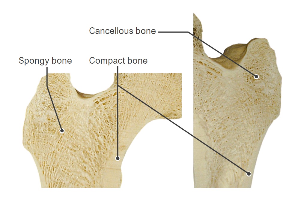
Haversian canals are located in which type of bone?
- Compact bone
- Spongy bone
- Cartilaginous bone
- Fibrous bone
Which of the following options best describes the system through which osteocytes obtain cell nutrition?
- Lacunar-canalicular pore systems filled by osteocyte processes
- Diffusion from the vascular endosteum
- Diffusion from the outer layer of the periosteum
- Diffusion from the inner layer of the periosteum
- The majority of osteocytes are dead and do not require nutrition.
Which of the following options ONLY includes components of an osteon?
- Concentric lamellae, osteocytes, canaliculi, Haversian canals
- Bony spicules, concentric lamellae, osteocytes, interstitial lamellae
- Endosteum, bony spicules, Haversian canals, osteocytes, periosteum
- Osteocytes, concentric lamellae, Haversian canals, periosteum
- Interstitial lamellae, osteocytes, concentric lamellae, Haversian canals
Which of the following refers to a series of microscopic tubes in the cortical bone for the passage of blood vessels and nerves?
- Haversian canals
- Bony spicules
- Concentric lamellae
- Interstitial lamellae
Osteocytes are embedded in which of the following?
- Mineralized bone matrix
- Haversian canals
- Noncalcified matrix
- Endosteum
Which of the following best describes the histology of a cross-section of Haversian canals?
- Circular openings at the center of osteons
- Dense structures at the center of osteons
- Circular openings surrounding osteons
- Dense structures surrounding osteons
- Haversian canals are not readily distinguished under a microscope.
Author of lecture Osteon

Geoffrey Meyer, PhD
Customer reviews
5,0 of 5 stars
| 5 Stars |
|
5 |
| 4 Stars |
|
0 |
| 3 Stars |
|
0 |
| 2 Stars |
|
0 |
| 1 Star |
|
0 |
