Playlist
Show Playlist
Hide Playlist
Osteoarthritis: Risk Factors and Radiography

-
Slides Osteoarthritis.pdf
-
Reference List Rheumatology.pdf
-
Download Lecture Overview
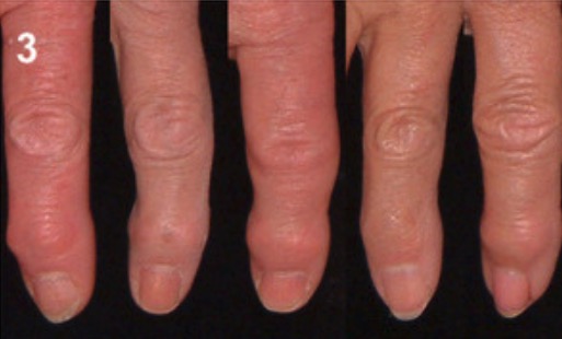
00:00 So what causes osteoarthritis? this one's easy. 00:05 Despite being the most common, chronic, degenerative joint disease, afflicting nearly everybody over the age of 50, the etiology of osteoarthritis remains completely unknown. 00:16 We do, however, at least know some of its risk factors. 00:19 Anyone over the age of 65 has osteoarthritis and oftentimes it can start considerably earlier. 00:25 Female gender is a risk factor, any family history, prior joint trauma and certainly obesity by virtue of excess wear and tear on those joints. 00:34 In addition, osteoarthritis can be secondary to other disease processes like diabetes, hemochromatosis, acromegaly and even gout can cause osteoarthritic types of manifestations. 00:46 So, let's go back to our case for a moment and look for some key features. 00:50 We have an acute-on-chronic picture, very typical for osteoarthritis. 00:54 Worse after activity but morning stiffness lasting for less than 15 minutes. 00:59 Also, typical of osteoarthritis. 01:02 The swollen joint can happen as an acute synovitis on a backdrop of chronic osteoarthritis. 01:08 It's not common but it does certainly happen. 01:11 Bony enlargement of the knees due to these osteophytic growths around the pes is common especially on radiographs. 01:17 and the fact that you have tenderness on the medial joint line also supports that condition. 01:23 So, to recap: Classic features of osteoarthritis are those we had in our case. 01:28 A chronic disease with periodic flares, typically precipitated by increased activity. 01:33 You're also looking for asymmetric symptoms and with inexorable progression osteoarthritis of some joints can literally bring many people to their knees. 01:43 See what I did there? Now, it has a predilection for certain joints. 01:48 Like the knees, the hips, the lumbar spine and of course shown here on the left, the proximal interphalangeal and the distal interphalangeal joints of the fingers. 01:58 Classic physical exam findings as I alluded to are gonna be bony enlargement, a limited range of motion, varus deformity which means being somewhat bowlegged, crepitus by palpating the knee while moving it and of course, joint-line tenderness as I've mentioned. 02:14 So let's take a look at some of the common radiographic features of osteoarthritis. 02:18 The picture on the left here is a normal radiograph of a knee and the one on the right is somebody with severe osteoarthritis. 02:25 What you can see right off the bat, the first distinguishing feature is the joint space narrowing, particularly in the medial compartment of the knee. 02:32 In addition, you can see evidence of subchondral sclerosis which is that hyperlucent areas just on the tibial plateau. 02:39 There's evidence of subchondral cyst in there as well and osteophytes basically broaden the size of the knee relative to the size of the tibial plateau.
About the Lecture
The lecture Osteoarthritis: Risk Factors and Radiography by Stephen Holt, MD, MS is from the course Non-Autoimmune Arthritis.
Included Quiz Questions
Which of the following is NOT commonly seen on an X-ray of osteoarthritic joints?
- Subchondral erosions
- Joint space narrowing
- Osteophytes
- Subchondral cysts
- Subchondral sclerosis
Author of lecture Osteoarthritis: Risk Factors and Radiography

Stephen Holt, MD, MS
Customer reviews
5,0 of 5 stars
| 5 Stars |
|
5 |
| 4 Stars |
|
0 |
| 3 Stars |
|
0 |
| 2 Stars |
|
0 |
| 1 Star |
|
0 |
