Playlist
Show Playlist
Hide Playlist
Orbital Cellulitis and Preseptal Cellulitis: Pathology & Clinical Presentation

-
Slides preseptalorbitalcellulitis Pediatrics.pdf
-
Download Lecture Overview
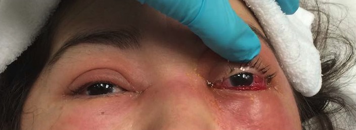
00:00 In this lecture, we're going to discuss Orbital Cellulitis and Preseptal Cellulitis. Basically, a child who comes in with an infected red eye. It's important to distinguish between these 2 things because we treat them differently. Bear with me here. Imagine if you will a bunch of firefighters around one of those old trampolines and they're underneath the building and they're screaming "jump, jump" to the poor person who is up on the third floor who they want to save. Let's think about that trampoline they're holding and let's imagine that we have this trampoline but that center spot where the red dot is, we cut it out so there's a little hole. So here is that trampoline with the little hole in it. Now let's drop a ball into the hole but the ball is bigger than the hole so it sits in place. There's the ball. This is essentially the eye, the ball is the eyeball and the green part is the septum and that septum is holding the eye in place just like that ball is being held up. So here's a side view of an eyeball. That ocular septum, the orbital septum is coming down and around the eye and there's an extension so that that eye is firmly held into the head. 01:26 However, that septum serves more than just a structural role. It prevents bacteria from growing in 1 direction or the other. So infections in front of the septum will be different than the ones behind the septum. That septum is acting as a physical structure that holds the eye in place but also a barrier for infectious organisms. So if we look at this eye again, infections outside the eye we call preseptal or before the septum cellulitis. These infections are mostly from the skin and the number one organisms are <i>Staph</i> and group A <i>Strep</i>. However, if an infection is here behind the septum, this is orbital cellulitis and these infections don't come from the skin most commonly most commonly they come from the sinuses because the sinus is a paper-thin wall away from the orbit. So, let's look then at the differences between these 2 infections. Orbital cellulitis is a mixed floral infection from the sinuses. It includes gram positives, gram negatives and even anaerobes and it requires broad-spectrum antibiotics to kill those bacteria. Also, there is a vision risk. This is an emergency. Preseptal cellulitis is usually group A <i>Strep</i>. We usually can treat it with narrow-spectrum antibiotics like a first generation cephalosporin and it rarely spreads into the eye if we treat it correctly. So, this 18-month-old is coming in to see you and he has a swollen eye. How do I tell if it's preseptal cellulitis and I require only a narrow-spectrum first generation cephalosporin or it's a postseptal infection called orbital cellulitis and any broad-spectrum antibiotics and I have to worry about this patient? How do I tell the difference? Well, there are number of ways we can tell the difference and here's all the symptoms and whether it's likely in preseptal or orbital. Orbital cellulitis can present with just about everything but preseptal cellulitis, it's a little clearer. Let's look at these in 2 groups. In preseptal cellulitis, patients will have eyelid swelling, redness and discharge. They can have that also in orbital cellulitis and they will have a normal pupillary response which is usually the case in orbital cellulitis but certainly if you see an abnormal pupillary response you should worry about that patient but preseptal cellulitis does not present with diplopia, with abnormal eye movements, with pain with eye movements, or with proptosis. If you see any of these things, you should presume this is orbital cellulitis. So, let's talk a little bit about diplopia. We need to understand where these sinuses are and why these patients with orbital cellulitis but not preseptal cellulitis get diplopia and pain with eye movements. So here's a little child who's presenting with a red eye and this child has an infection of their ethmoid sinus. Remember that ethmoid sinus is just medial to the eye, which means that when that infection broke through that paper-thin bone, it entrapped the medial rectus muscle of this patient's eye. So now we'll ask this patient to look around. 05:01 I asked them to look to the side towards their affected ethmoid sinus, they should be doing okay but when I asked them to look away from the infected sinus, that infected medial rectus muscle is entrapped. They can't look out. So if I see a patient who looks like this, I can say very comfortably this is probably an orbital cellulitis and I'll bet you it's from an ethmoid sinusitis. 05:32 Now, here's a little boy coming to see us. He has also got a red eye but this little boy has infection of his maxillary sinuses. So now this infection is going to escape upward into the inferior rectus muscle just below the eye. Let's have this boy look around. He can look to the left. Right? He can look to the other side. Right? He's doing okay with lateral movement. When we asked him to look down, he's fine but when we asked him to look up his inferior rectus muscle is entrapped and he has a dysconjugate gaze. It hurts for him to look up and you can see that his eye doesn't want to go that way.
About the Lecture
The lecture Orbital Cellulitis and Preseptal Cellulitis: Pathology & Clinical Presentation by Brian Alverson, MD is from the course Pediatric Infectious Diseases. It contains the following chapters:
- Pathology of Cellulitis
- Clinical Presentation of Cellulitis
Included Quiz Questions
Which of the following is true about preseptal cellulitis?
- It rarely spreads into the eye.
- You should treat the patient with broad spectrum antibiotics.
- It presents with pain on eye movement.
- It is caused by anaerobes.
- It is usually caused by a preceding sinusitis.
Which of the following is seen in BOTH preseptal and orbital cellulitis?
- Eyelid swelling, redness and discharge
- Diplopia
- Abnormal eye movement
- Pain with eye movement
- Proptosis
A child has an infection of the ethmoid sinus causing an orbital cellulitis. Which extraocular muscle can become entrapped causing a disconjugate gaze?
- Medial rectus
- Lateral rectus
- Levator palpebrae superioris
- Superior rectus
- Inferior rectus
A patient has a disconjugate gaze and pain in his left eye when looking up. Assuming he has an orbital cellulitis due to bacterial spread, which sinus is the likely source of infection?
- Maxillary sinus
- Frontal sinus
- Ethmoidal sinus
- Mandibular sinus
- Sphenoid sinus
Author of lecture Orbital Cellulitis and Preseptal Cellulitis: Pathology & Clinical Presentation

Brian Alverson, MD
Customer reviews
5,0 of 5 stars
| 5 Stars |
|
2 |
| 4 Stars |
|
0 |
| 3 Stars |
|
0 |
| 2 Stars |
|
0 |
| 1 Star |
|
0 |
Understanding that orbital cellulitis typically comes from sinus infections, whereas preseptal usually comes from the skin is helpful.
I've seen several preseptal cellulitis in children but the attendings could never explain this disease as well as it has been done in this lecture. Thumbs up!
