Playlist
Show Playlist
Hide Playlist
Optic Nerve


-
Slides 16 Human Organ Systems Meyer.pdf
-
Reference List Histology.pdf
-
Download Lecture Overview
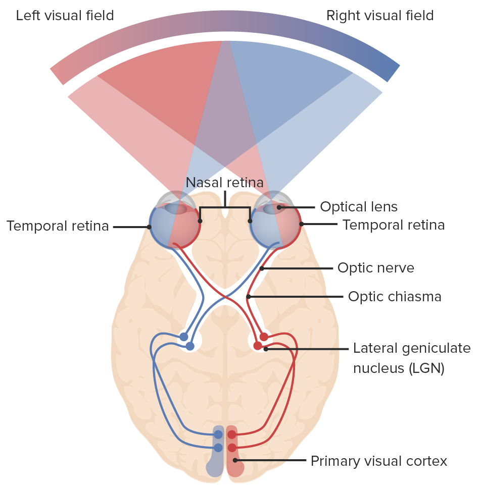
00:00 Let me just point out the structure of the optic nerve, in brackets they are put tract for the reason that I explained earlier in the lecture. On the left-hand side, you can see the optic nerve with a very characteristic central artery in the middle. And the purple stain component you see there are all the axons and they are supporting cells. 00:27 Those axons are projecting from the ganglion cells in the retina at the optic disc, which does not have any photoreceptors in it, so that's our blind spot. 00:38 On the outside of that image that section, you can see a dense connective tissue layer. That is the dura, one of the outer meninges of the brain protecting the brain and spinal cord. Then you see an artificial space, a white space and then a thin line, normally that thin line is up against the dura and you have a space that that thin line is the arachnoid layer and underneath that, that space is real. That is a subarachnoid space. Ond on the right-hand side, a higher magnification you then have the pia directly in contact with the surface of the ganglion axons unmyelinated nerve fibres and that is the pia component and that actually extends into the bundles of unmyelinated axon and supports them. So the optic nerve is surrounded by meninges, not the epiphery and endomysium, you find around peripheral nerve which is why I keep saying it should be called a nerve tract rather than just a straight peripheral nerve, so optic tract is a better name for the structure. 02:03 The supporting cells you can just see little nuclei and they are going to be glial cells.
About the Lecture
The lecture Optic Nerve by Geoffrey Meyer, PhD is from the course Sensory Histology.
Included Quiz Questions
In its final anatomical position before entering the optic disk, the central retinal artery lies in what position relative to the optic nerve?
- Within the optic nerve
- Inferior to the optic nerve
- Superior to the optic nerve
- Lateral to the optic nerve
- Medial to the optic nerve
Which of the following layers of protective tissue does NOT cover the optic nerve?
- Periosteum
- Arachnoid
- Pia
- Subarachnoid space
- Dura
Author of lecture Optic Nerve

Geoffrey Meyer, PhD
Customer reviews
5,0 of 5 stars
| 5 Stars |
|
5 |
| 4 Stars |
|
0 |
| 3 Stars |
|
0 |
| 2 Stars |
|
0 |
| 1 Star |
|
0 |
