Playlist
Show Playlist
Hide Playlist
Omphalocele

-
Slides GIP Omphalocele.pdf
-
Reference List Pathology.pdf
-
Download Lecture Overview
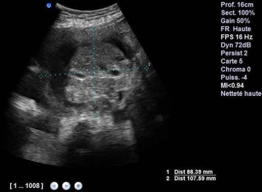
00:01 This talk is about Omphalocele and it may seem like omphalocele and gastroschisis are the same thing. 00:07 Not quite, they have a little bit different etiologies in terms of the developmental biology. 00:14 So omphaloceles are defects of the anterior abdominal wall. 00:17 This is at the base of the umbilicus. 00:19 And we'll see in a minute exactly why that happens. 00:23 As opposed to gastroschisis, the viscera in omphalocele is covered by the peritoneum, and by amniotic membranes, So it will look quite different, both grossly as well as by ultrasound. 00:36 So the epidemiology of omphalocele. 00:39 It has a slightly higher prevalence than gastroschisis about 1 in 5,000 births, and occurs for reasons that are obscure. 00:47 Boys more frequently than girls. 00:50 The maternal risk factors include very young age for a mom or much older moms. 00:58 And it can be part of syndromes associated with various chromosomal abnormalities. 01:04 The pathophysiology really has to do with the failure of the midgut to return to the abdominal cavity at 10 weeks gestation. 01:11 As we'll see, a lot of the bowel early on kind of protrudes into the yolk sac, and then develops in that location and then returns to the abdominal cavity. 01:22 If we don't do that appropriately, we don't get the abdominal folds to seal and it leaves a midline defect with bowel herniated out. 01:32 So here's our baby at 7 weeks of gestation. 01:35 And you can see going out into the umbilical cord region into the yolk sac region, we can see loops of bowel, that's completely normal. 01:45 However, by about 12 weeks, that's all returned back into the abdominal cavity. 01:51 And there will be some folding and other things that go on. 01:53 We'll talk about that when we talk about disorders of malrotation. 01:58 However, in the setting of omphalocele, we don't get that to return. 02:03 It stays outside. 02:05 And we have loops of bowel surrounded by a membrane that stick out of the midline right along where the umbilicus normally would be. 02:14 The clinical presentation. 02:16 You can see here at birth and herniation of intestines through an umbilical ring and it's completely surrounded by a membrane. 02:24 So that amniotic membrane and the peritoneum allow this to be somewhat protected. 02:29 So you don't have as much fluid loss or as much of a risk of infection as we would with gastroschisis. 02:37 The diagnosis and monitoring. 02:39 So ultrasonography is again, our best friend in trying to diagnose whether omphalocele is present. 02:45 In most cases, we pick that up very early on in pregnancy. 02:50 And it's part of the routine prenatal screening. 02:53 There may be other abnormalities. 02:55 And remember that there may be other genetic defects associated with this, so we want to be screening not just for omphalocele but for other possible abnormalities. 03:05 Again, shown here in ultrasonography, we have a big loop, several loops of bowel sitting outside of the abdominal cavity, and they do have a membrane around them. 03:15 So you can actually distinguish this from gastroschisis, just on the ultrasound. 03:22 Okay, so what do we do about omphalocele? Let's go to management and prognosis. 03:29 Because this loop of bowel outside of the abdomen can actually compromise respiratory excursion by limiting diaphragmatic movement, you may have or the baby may have lung hypoplasia. 03:42 And if this is unrecognized can lead to respiratory distress early on after delivery. 03:48 We have to be aware of that. 03:50 We also need to avoid just poking the thing back in and sewing it back up. 03:54 Those loops of bowel are prone to perforation. 03:57 So we have to be very careful and avoid just a simple manual reduction. 04:02 For small to medium size defects. 04:05 And there's normal lung activity: there's normal lung function, then the primary repair where we cut back the membrane and carefully reinsert the bowel back into the abdomen. 04:20 Close the defect, and we're good to go. 04:24 And with that, we conclude our discussion on omphalocele and gastroschisis developmental defects of the abdominal wall.
About the Lecture
The lecture Omphalocele by Richard Mitchell, MD, PhD is from the course Congenital Gastrointestinal Tract Disorders.
Included Quiz Questions
Where is the location of the defect in an omphalocele?
- The base of the umbilicus
- Lateral to the umbilicus
- Superior to the umbilicus
- Right upper quadrant of the abdomen
- Left upper quadrant of the abdomen
What is the pathophysiology of an omphalocele?
- Failure of midgut to return to abdominal cavity around 10 weeks gestation
- Failure of foregut to return to abdominal cavity around 10 weeks gestation
- Failure of hindgut to return to abdominal cavity around 10 weeks gestation
- Failure of midgut to return to abdominal cavity around 12 weeks gestation
- Failure of hindgut to return to abdominal cavity around 8 weeks gestation
Author of lecture Omphalocele

Richard Mitchell, MD, PhD
Customer reviews
5,0 of 5 stars
| 5 Stars |
|
5 |
| 4 Stars |
|
0 |
| 3 Stars |
|
0 |
| 2 Stars |
|
0 |
| 1 Star |
|
0 |
